Full transparency
The pre-operative assessment for refractive surgery
The pre-operative assessment is the step that comes before any laser refractive surgery. This consultation allows me to determine whether your eyes can be operated on and to choose the right technique. I also give you a precise idea of the expected result. For your part, you explain your plans and ask your questions. You make your decision fully informed.
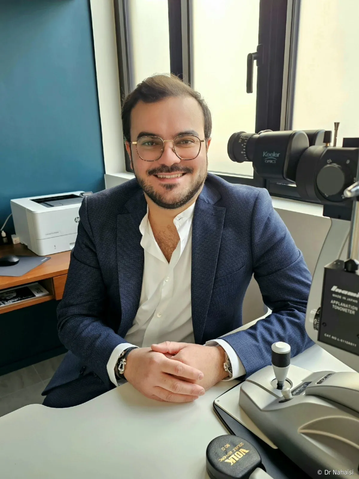
Dr Steven Nahaisi
Updated on 15 October 2026 · 26 min read
A single consultation is almost always enough. No drawn-out process, no back-and-forth. You leave with a clear answer.
On this page10 sections
- 02WHY A PRE-OPERATIVE ASSESSMENT
- 03HOW THE ASSESSMENT WORKS IN PRACTICE
- 04THE INTERVIEW · UNDERSTANDING YOUR PLANS
- 05THE TESTS PERFORMED
- 06WHAT I LOOK FOR · THE CRITERIA FOR SUITABILITY
- 07MY VIEW ON SUITABILITY FOR SURGERY
- 08AT THE END OF THE ASSESSMENT
- 09CONSULTATION FEE
- 10FREQUENTLY ASKED QUESTIONS
- 11BOOK AN APPOINTMENT
WHY A PRE-OPERATIVE ASSESSMENT
What is the assessment before laser eye surgery for?
Laser refractive surgery is tailored to each eye. Every cornea has its own shape, thickness and curvature. Every patient comes with their own lifestyle and expectations. The pre-operative assessment makes it possible to tailor the procedure to both of these dimensions.
This assessment has four objectives.
Understanding your plans and your lifestyle
Before the measurements, I listen. Why you are coming today, what you expect from surgery, how you live with your glasses or contact lenses, what you are afraid of. The discussion guides the choice of technique.
Checking that your eyes are suitable for surgery
Laser surgery is possible for most patients. But there are situations where it is not: a cornea that is too thin, signs of weakness, a correction beyond the limits of the laser. My role is to detect these beforehand.
When laser surgery is not the right option, I say so clearly. I would rather not operate than take the slightest risk with your eyes.
Choosing the most suitable technique
LASIK or TransPRK for most cases, SMILE or presbyopia strategies depending on the indication. No technique is better than the others in absolute terms; there is the one that matches your eye and your correction. The assessment tests make it possible to decide on objective criteria. Your lifestyle and your professional or sports constraints also guide the choice.
Anticipating the result and screening for anything that could be a problem
The assessment also lets me give you a precise idea of the expected result. This is when I screen for the most subtle signs of weakness, early keratoconus being the most important example. My hospital experience in corneal surgery, with more than 350 corneal transplants, taught me to spot them.
HOW THE ASSESSMENT WORKS IN PRACTICE
How long does the pre-operative assessment before LASIK take?
The assessment takes place in a single session. Allow a good hour on site, enough time to talk together, carry out all the tests, explain the recommended technique to you, and answer all your questions.
No need for any other intermediate consultations. If surgery is approved, the next appointment is the operation itself.
Are drops used to dilate the pupils?
Not in most cases. The fundus (back of the eye) examination is done with retinal photography, without dilation. You leave just as you came, and you can drive or go back to work right afterwards.
In some specific cases, I may need an additional test with drops (Skiacol, which temporarily paralyzes accommodation). This test measures your correction at rest, without interference from the focusing muscle. When it is needed, I explain it to you during the consultation and we arrange it either the same day or during a short second visit a few days later. It must then be done at least 3 days before surgery, to give your pupils time to return to their normal size.
Do I need to remove my contact lenses before the assessment?
Not systematically. Removing your contact lenses is not mandatory for the pre-operative assessment, but it can be useful in borderline cases, because contact lenses slightly change the shape of the cornea. Ideally, take them out on the morning of the consultation for more accurate measurements. If they were left in, the measurements are usable most of the time. And if they turn out to be borderline, we arrange a new set of measurements before surgery.
Not to be confused with removal before the operation itself: for surgery, soft contact lenses are removed 48 hours before the procedure, so that the cornea is examined and operated on in its natural state. For rigid lenses, it is decided case by case: I check with repeated topographies that the curvature of the cornea is stable.
Do I need a referral letter from another ophthalmologist?
No. No referral letter, no doctor's note, no prior tests. You can book an appointment directly, through Doctolib, by phone or by email. I have all the necessary equipment on site, at the Institut Voltaire as well as at the satellite practices.
What should I bring?
Not much. Come with your usual glasses or contact lenses. If you have glasses prescriptions from recent years, they help me check whether your correction has changed, but they are not mandatory. And if you have already had corneal tests (topographies, for example), bring them; otherwise, I will redo everything on site.
No fasting, no preparation
No special preparation. You eat normally and take your medication as usual. The assessment requires neither fasting nor stopping any treatment.
THE INTERVIEW · UNDERSTANDING YOUR PLANS
The consultation starts with a conversation
The assessment starts with time to talk. Understanding who you are, why you are coming and what you expect from this surgery is as necessary to me as measuring your cornea. The chosen technique and the correction strategy are largely built around this conversation. So is the pace.
I often say that I don’t operate on eyes, I operate on patients.
Why now: your motivation and your expectations
I start with the obvious questions: what brings you here, and why now? Being tired of glasses, contact lens intolerance, a life plan, a sport, a demanding job, a cosmetic reason. All of these reasons are valid. I try to understand what you really expect from surgery. Complete independence from glasses? Sharp distance vision? Being able to read with nothing on your nose?
I weigh this goal against what surgery can actually achieve for your eyes. Refractive surgery has its limits. My role is to match, as far as possible, your plans with what the technique allows, and to tell you clearly when the gap is too wide.
Your daily life: visual habits, work, driving
How many hours do you spend in front of a screen? Do you read a lot? Do you drive a lot, especially at night? These answers matter. Prolonged screen work makes post-operative eye strain more likely in the first weeks, and sometimes leads to prescribing rest glasses after surgery. Intensive night driving calls for caution regarding the management of halos and the correction strategy, particularly in presbyopic patients.
As for contact lenses, I look at your real comfort, beyond mere tolerance. How long do you wear them each day? Do you suffer from discomfort at the end of the day, dryness, red eyes? These signals weigh in the decision, particularly when there is a tendency to dry eye.
Sports and profession, when lifestyle drives the technique
This factor directly influences the choice of technique.
For combat sports (boxing, MMA, krav maga), intense contact sports, law enforcement, the military, firefighters, in short any situation where your eye is exposed to a risk of trauma, I often favor TransPRK over LASIK. TransPRK does not create a corneal flap and offers additional mechanical safety in case of a blow to the eye. In boxing, with gloves, a LASIK flap is not a problem: it is mostly a question of peace of mind.
LASIK remains excellent for the vast majority of patients, athletes included. The risk of the flap shifting in everyday life is very low. But for profiles exposed to repeated trauma, I sometimes prefer to remove this variable.
For visually demanding professions (pilots, surgeons, precision work, night work, artistic professions), expectations in terms of quality of vision are often higher than average. We discuss this in detail, and the surgical strategy is adjusted.
Your anxieties or fears, a topic in their own right
Refractive surgery is elective surgery. No one has to have it. It is a choice, and that choice brings a lot of apprehension. Will it work? Does it hurt? What if something goes wrong? I hear these questions every day, and I take the time to answer them one by one.
I prefer to explore these fears with you. Some patients are afraid of the laser itself, others of the operating room, others of the result, and others still of a specific event they have heard about. Understanding exactly what worries you makes it possible to address the right point. When a patient needs it, I offer two consultations rather than one, or a prior contact lens trial to test the correction strategy (particularly for presbyopic patients considering monovision). There is no rush.
Beyond the consultation, you can write to me at any time, before as well as after the operation. I answer my patients personally.
THE TESTS PERFORMED
What tests are done during the assessment before refractive surgery?
Once the discussion is over, we move on to the measurements. The assessment combines a general eye examination with a targeted assessment of the cornea and optical aberrations. Here, in order, is what we do.
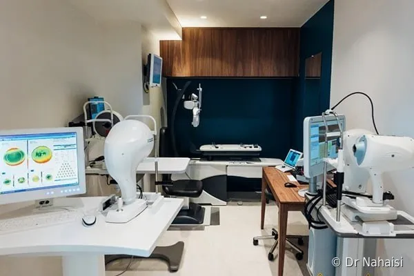
The diagnostic equipment at the Institut Laser Ophtalmologique Voltaire (Voltaire Laser Eye Institute). All the assessment tests are performed on site, with complementary devices: corneal topographers, anterior segment OCT, aberrometer, retinal camera. So you complete the whole assessment in a single consultation, without being sent from one center to another. © Dr Nahaisi.
1. Refraction and visual acuity
We measure your precise correction (sphere, cylinder, axis for each eye) and your visual acuity with and without correction. This measurement is compared with your current prescription and your history. A minimal variation of 0.25 or 0.50 diopters between two examinations is normal. It is a natural fluctuation of the eye, with no real change in your correction.
2. Corneal topography, the map of your cornea
This is the central test of the assessment. Corneal topography measures the curvature of your cornea point by point, on its front and back surfaces. It produces a color map that looks like a topographic relief map, hence its name.
At the Institut Voltaire, I use several complementary topographers to cross-check the measurements and get a complete picture. The more regular and symmetrical the cornea, the more suitable it is for laser treatment. Conversely, asymmetries, even subtle ones, can be a sign of early keratoconus and contraindicate surgery.
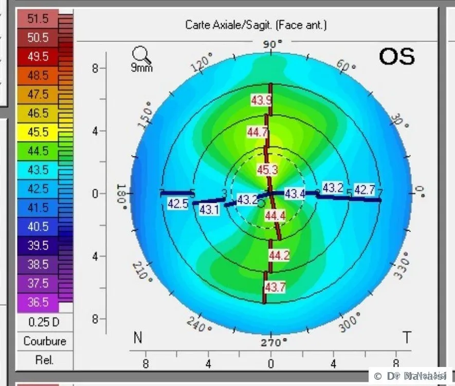
Example of a normal corneal topography (axial curvature map). Warm colors correspond to the steepest areas, cool colors to the flattest areas. Here, the cornea is regular and symmetrical: the pattern is balanced around the center, with no localized thinning or bulging. This is the profile of a cornea suitable for laser surgery. A suspicious cornea would instead show asymmetry, an off-center “bow tie” or an isolated bulging area, all signs of early keratoconus. © Dr Nahaisi.
3. Pachymetry, the thickness of your cornea
Pachymetry measures the thickness of your cornea, in microns. A cornea measures 525 microns on average. For LASIK, the standard threshold is a pre-operative thickness of 500 microns. For TransPRK, thinner corneas can be treated.
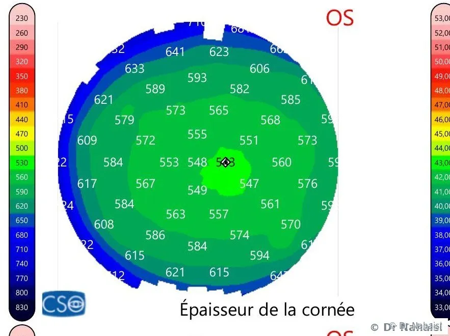
Corneal thickness map (pachymetry) measured with anterior segment OCT (MS-39). The cornea is not the same thickness everywhere: it is thinnest in the center (here about 545 µm, in green) and gradually thickens toward the periphery (600 to 650 µm, in blue). This distribution has a direct consequence for surgery. In nearsighted eyes, the laser sculpts the center, that is, the thinnest area, which makes central corneal thickness and the residual bed particularly decisive. In farsighted eyes, the laser works in the periphery, where the cornea is thickest, but the correction is then limited mainly by the maximum curvature that can be given to the cornea. The factors that limit surgery are therefore not the same depending on the error to be corrected. © Dr Nahaisi.
But thickness alone is not enough. A regular cornea at 490 microns can be more suitable for LASIK than a cornea at 520 microns with irregularities. What matters is the whole picture: thickness, regularity, curvature, the correction to be removed, and the residual posterior stromal bed.
4. Epithelial thickness, a key test for detecting weakness
The epithelium is the surface layer of the cornea, its “skin”. It is about 50 microns thick. Mapping it point by point provides valuable information for keratoconus screening: localized thinning of the epithelium over a bulging area is an early sign of corneal asymmetry. Conversely, a regular epithelium, or even inferior thickening, is a sign of good suitability for surgery.
This test, rarely performed in general ophthalmology, is a systematic part of the pre-operative assessment at the Institut Voltaire. It carries a lot of weight in early screening.
5. Aberrometry, your eye is not just an average value
When we say an eye has “-3 diopters”, that is an average value. Your eye is actually a complex surface, made up of an infinite number of points, each with its own correction: points at -2.90, others at -3.10, and so on. This fine distribution is called the aberrometry map.
The laser, for its part, treats the cornea point by point, adapting to this actual map. The Schwind platform I operate on uses the aberration-free profile for this, which customizes the treatment to your eye. The result is more precise than what correcting an average value with glasses allows.
Aberrometry is measured during the assessment, then repeated on the day of the procedure to feed the most recent data into the laser.
Learn more about aberrometry and customized treatment
6. Pupil measurement
We measure the size of your pupil in daylight and in low light. This data is important for two reasons. First, it makes it possible to calibrate the optical zone of the laser treatment so that it amply covers the pupil even when dilated, which reduces the risk of halos at night. Second, a large pupil in scotopic (low-light) conditions is a factor to include in the surgical strategy, particularly for patients who drive a lot at night.
7. Ocular surface examination
We assess the quality of your tear film and the surface of your cornea. Pre-existing dry eye does not contraindicate surgery, but it can influence the choice of technique and guides post-operative care. An allergic tendency, a history of dry eye, prolonged screen work: all factors I take into account.
8. Fundus examination
Usually performed without drops, by retinal photography. It makes it possible to check that the retina is healthy. In highly nearsighted patients, whose retina is thinner and more stretched, this examination is particularly important. Refractive surgery corrects the cornea but does not change the structure of your retina. Regular retinal follow-up remains necessary after the operation.
9. Intraocular pressure measurement
A quick, systematic test to screen for ocular hypertension or possible early glaucoma.
WHAT I LOOK FOR · THE CRITERIA FOR SUITABILITY
How do I know if my cornea is suitable for LASIK?
Suitability for surgery is not just about corneal thickness, contrary to a widely held idea. It depends on several parameters, and it is their combination that determines the decision.
There are in fact dozens of statistical formulas assessing the risk of post-operative ectasia (the feared adverse effect of LASIK), each giving more or less weight to one parameter or another. My role as a surgeon is to review all of this data comprehensively, and to make an informed diagnosis of suitability.
Total corneal thickness
The standard threshold for LASIK is 500 microns pre-operatively. Below that, we tend to opt for TransPRK, which does not require a flap and better preserves residual thickness. Thickness alone does not settle the matter, however: a thin but regular cornea may be suitable, whereas an average but irregular cornea will not be.
Corneal regularity, screening for keratoconus
Keratoconus is the main contraindication to refractive surgery. This progressive deformation of the cornea, which stretches and bulges into a cone, can be triggered or worsened by LASIK in a predisposed patient. It must therefore be screened for before any procedure.
Advanced forms are easy to recognize; early, subclinical forms are less so. Subtle asymmetry, localized thinning, epithelial irregularity: all subtle signs that require a trained eye. My hospital background in corneal surgery (Hôtel-Dieu with Prof. Bourges, Fondation Rothschild with Prof. Gabison and Prof. Gatinel, then former Chief Resident (Chef de Clinique) at Cochin Hospital) trained me to detect them, and to tell truly suggestive signs from false alarms.
The residual posterior stromal bed
This is the thickness of intact cornea left behind the laser treatment. In LASIK, about 110 microns are taken up by the flap, then an additional 15 microns per diopter of myopia corrected. What remains is the residual stromal bed. It is what guarantees the long-term stability of your cornea.
Animated simulator of the residual stromal bed. In LASIK, the femtosecond laser cuts a 110 µm flap attached by a hinge, the excimer sculpts only the central optical zone (≈ 75 µm), then the flap is laid back flat. The residual bed is about 365 µm. In TransPRK, the laser removes the central epithelium (≈ 50 µm, which regenerates) then sculpts the stroma without creating a flap: the residual bed remains higher, about 425 µm. For the same correction, TransPRK thus preserves around sixty microns more, because it does not sacrifice a layer of stroma in a flap. © Dr Nahaisi.
Standard recommendations set the minimum threshold at 280 microns. Some studies go as low as 250 microns, but this is a reduced margin that assumes all the other parameters are perfectly reassuring. For my part, I take a more cautious approach, at 300 microns, to ensure an additional safety margin.
I do this calculation for each eye when preparing the treatment. The laser software (below) simulates the ablation and displays the expected residual stromal bed (“RST”, residual stromal thickness), which I systematically check before approving surgery.
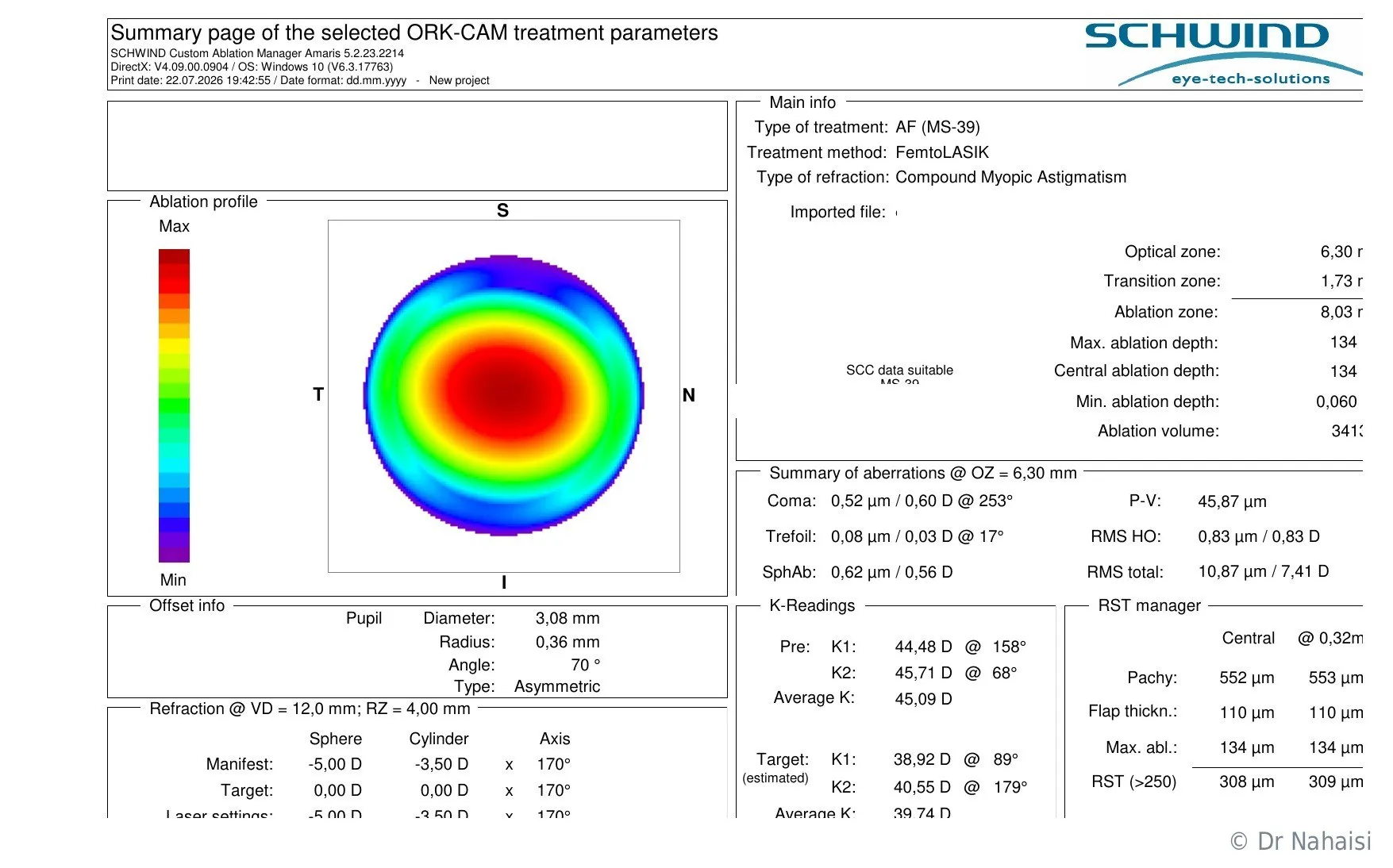
Treatment plan for a nearsighted, astigmatic eye (anonymized example). On the left, the ablation profile: the laser removes tissue in the center, in red, and barely touches the periphery. On the right, the “RST manager” calculates the residual stromal bed from the corneal thickness (552 µm), the flap thickness (110 µm) and the ablation depth (134 µm). 308 µm remain, above the safety threshold. I do this check for every patient before approving surgery.
When the expected residual bed would be insufficient with LASIK, because the cornea is thin or because the correction is high, TransPRK becomes the technique of choice. For very high corrections beyond -10 diopters, the phakic implant (ICL) is also an option to consider, with referral to a specialized colleague.
Corneal curvature, anterior and posterior keratometry
We measure the curvature of your cornea, both on its front surface (anterior keratometry, as mean and maximum values) and on its back surface (posterior keratometry). A marked asymmetry between the two surfaces can be a sign of weakness.
The starting curvature also determines the possible range of correction. A cornea that is too flat in a nearsighted patient, or too steep in a farsighted patient, limits the laser’s room for maneuver: in the first case, because an already flat cornea would have to be flattened further; in the second, because it would exceed reasonable limits of post-treatment steepening. This point is detailed on the dedicated pages.
Eye rubbing
Chronic rubbing is one of the main causes of corneal deformation and acquired keratoconus. When the topography shows a sign of weakness, I systematically try to find out whether there is a risk behavior: rubbing in the morning on waking, allergic itching, sleeping on your stomach with your head in the pillow, a hand or elbow pressed under the eye during the night.
Identifying and changing these behaviors is sometimes enough to stabilize a fragile cornea and consider surgery after a few months of monitoring.
Summary: the parameters analyzed and what they determine
| Parameter | Test | What it determines |
|---|---|---|
| Total thickness | Pachymetry | Feasibility of LASIK; TransPRK if thin |
| Regularity | Corneal topography | Keratoconus screening, asymmetry, overall suitability |
| Epithelial thickness | Epithelial mapping | Early detection of corneal irregularities |
| Curvature (front and back surfaces) | Keratometry / topography | Ectasia risk; treatment limits |
| Posterior stromal bed | Calculated from thickness + flap + ablation | Long-term stability of the cornea |
| Aberration map | Aberrometry | Programming of the customized treatment |
| Pupil size | Pupillometry | Optical zone calibration; risk of halos at night |
| Ocular surface | Slit-lamp examination | Dryness, allergic tendency, choice of technique |
| Retina and optic nerve | Retinal photography, OCT if needed | Screening for associated disease |
MY VIEW ON SUITABILITY FOR SURGERY
How I decide whether a cornea is suitable for surgery
I often say in consultation that I look for the slightest reason why a cornea would not be suitable for surgery. That is how I work. Refractive surgery is elective surgery; my duty is to make it as safe as possible. That starts with a rigorous, uncompromising analysis.
When a case is clearly suitable, I tell you. When it is clearly contraindicated, I tell you too and refer you on. The most demanding work concerns borderline cases.
For complex cases: a joint opinion at the Institut Voltaire
At the Institut Laser Ophtalmologique Voltaire, I work with my colleagues Dr Camille Rambaud and Dr Romain Nicolau, who are also specialized surgeons. Every week we hold joint discussions on borderline cases. Complex cases are analyzed by several of us, and sensitive decisions are made together.
This three-way discussion provides a solid joint opinion. When we unanimously confirm suitability, you can go ahead with confidence. When we decide to wait, it is because caution requires it.
When laser surgery is not the right option
Sometimes the conclusion of the assessment is unfavorable. A cornea that is too thin and irregular, early keratoconus, a correction beyond the limits of the laser: all situations where I prefer not to operate. In these cases, I explain precisely why, and I present the alternatives to you.
For very high corrections beyond the limits of the laser, or for corneas that cannot withstand corneal surgery, the phakic implant (ICL) is often the right answer. I then refer you to a specialized colleague, while keeping control of coordinating your care if you wish.
When laser surgery is not possible: alternatives and referral
AT THE END OF THE ASSESSMENT
What happens at the end of the pre-operative consultation?
At the end of the consultation, I give you a clear answer: suitable or not, which technique, what expected result. If surgery is approved and you want to go ahead, you also leave with the complete surgical file.
The surgical file
It contains the appointment notice for surgery day, the prescription for the treatments to take before and after the operation, the informed consent form to sign, and the quote to send to your complementary health insurance to find out how much they cover.
See fees, quotes and complementary health insurance reimbursement
The date of the procedure
You can schedule your operation date at the end of the consultation, or later. No obligation. Some patients schedule their surgery in the following weeks. Others come back to me several months or several years later. Others never follow up, and that is perfectly legitimate. What matters is making your decision calmly and being ready.
In any case, I remain available. To get back to me, you can use Doctolib messaging, email or phone. I answer my patients personally, before and after surgery.
CONSULTATION FEE
How much does the pre-operative consultation for refractive surgery cost?
The pre-operative consultation is charged at the standard rate set by French national health insurance. At the 10th and 19th practices, there is no fee supplement. At the Institut Voltaire (11th), a €30 fee supplement applies.
What changes depending on the location is the amount you pay upfront.
| Location | Paid upfront | Details |
|---|---|---|
| Paris 10th (10e arrondissement), Centre Santé Plus | €0 | Standard rate, no fee supplement. Nothing to pay upfront (direct billing) with your carte Vitale and complementary health insurance. |
| Paris 19th (19e arrondissement), Cabinet Ophtavie | €0 | Standard rate, no fee supplement. Nothing to pay upfront (direct billing) with your carte Vitale and complementary health insurance. |
| Institut Voltaire (Paris 11th) | €50 | €30 fee supplement. Complementary health insurance reimburses at least €20, sometimes all of it depending on your policy. |
The consultation is reimbursed by French national health insurance at the standard rate. Complementary health insurance covers the rest depending on your policy.
Sometimes, at the 10th or 19th practices, I need an additional test that I only perform at the Institut Voltaire. I then refer you there to complete the assessment.
Frequently asked questions
Your questions about the pre-operative assessment for refractive surgery
How long does the pre-operative assessment before LASIK surgery take?
Allow about one hour on site. This includes the discussion with me about your expectations and your lifestyle, all the tests (refraction, topography, pachymetry, aberrometry, ocular surface examination, fundus examination), the explanation of the technique and time to answer all your questions. A single consultation is enough in the vast majority of cases. There is no second intermediate appointment before surgery.
Do I need a letter from an ophthalmologist to see a refractive surgeon?
No. No referral letter, no doctor's note, no prior tests. You can book an appointment directly through Doctolib, by phone or by email. I have all the equipment needed on site to carry out the complete assessment.
Are drops used to dilate the pupils during a LASIK pre-operative assessment?
Not in most cases. The fundus examination is done without drops, by retinal photography. You leave just as you came, and you can drive and resume your activities normally. In some specific cases, an additional test with drops (Skiacol) may be needed to measure your correction at rest. When that is the case, I explain it to you and we arrange it either the same day or during a short second visit.
Do I need to remove my contact lenses before a pre-operative assessment for refractive surgery?
Ideally, take out your contact lenses on the morning of the consultation for more accurate measurements. If you kept them in, the measurements are usable most of the time. For surgery, however, you will need to remove them 48 to 72 hours before the procedure.
Do I need to fast before a pre-operative assessment for laser eye surgery?
No. No preparation. You eat normally and take your medication as usual. The assessment requires neither fasting nor stopping any treatment.
What documents should I bring to a LASIK pre-operative assessment?
Not much. Come with your usual glasses or contact lenses. Your glasses prescriptions from recent years are useful for checking whether your correction has changed, but we manage without them if you don’t have them. If you have already had corneal tests (topographies), bring them; otherwise, I will redo everything on site.
Does the surgeon take my lifestyle into account during the pre-operative assessment for refractive surgery?
Yes. Before the tests, we discuss your daily life, your work, your sports, your driving, and what you expect from surgery. These factors directly guide the choice of technique: for a combat sport or an exposed profession, I often favor TransPRK over LASIK, for long-term safety reasons. For heavy night driving, I factor your pupil size and the risk of halos into the strategy. The discussion is an integral part of the assessment.
How do sports or profession influence the choice between LASIK and TransPRK?
For combat sports, contact sports, law enforcement, the military, firefighters, that is, any situation where the eye is exposed to a risk of trauma, I often favor TransPRK. This technique does not create a corneal flap and offers additional mechanical safety in case of a blow. LASIK remains excellent for the vast majority of patients, athletes included. The risk of the flap shifting in everyday life is very low. But for profiles exposed to repeated trauma, I sometimes prefer to remove this variable.
From what age can you have laser refractive surgery?
From age 18. The most favorable time is usually after age 22-23, when the correction has stopped changing. It is entirely possible to have the assessment earlier, and to operate before age 22 when the request is justified, by looking closely at stability over recent years and taking into account that the correction may still change.
How do I know if my cornea is too thin for LASIK surgery?
That is precisely the role of the assessment. The standard threshold for LASIK is a pre-operative thickness of 500 microns, but it is all the parameters together that count (regularity, curvature, expected residual bed). A regular cornea at 490 microns can be more suitable for LASIK than an irregular cornea at 520 microns. When LASIK is not the right option, TransPRK takes over for thinner corneas.
What is keratoconus and how is it screened for before LASIK?
Keratoconus is a progressive deformation of the cornea, which stretches and bulges into a cone. It is the main contraindication to LASIK, because the laser could worsen it. Screening relies on corneal topography (asymmetry, localized thinning), epithelial mapping, and analysis of both surfaces of the cornea. My hospital experience in corneal surgery, with more than 350 corneal transplants, trained me to detect its most subtle forms.
What happens if the pre-operative assessment shows that I am not suitable for laser eye surgery?
I explain precisely why, and I present the alternatives to you. For very high corrections beyond the reach of the laser, the phakic implant (ICL) is often the right answer. I then refer you to a specialized colleague. For corneas that are becoming weaker, it is sometimes possible to wait and stabilize the situation before considering surgery again. In any case, I would rather not operate than take the slightest risk with your eyes.
How is suitability decided in borderline cases in refractive surgery?
At the Institut Voltaire, I work with my colleagues Dr Camille Rambaud and Dr Romain Nicolau. When a case raises a question, we analyze it jointly, the three of us. This discussion provides a solid opinion, free of individual bias. When we unanimously confirm suitability, you can go ahead with confidence. When we decide to wait, it is because caution requires it.
Is the pre-operative assessment essential before laser refractive surgery?
Yes. No serious refractive surgery is done without a complete pre-operative assessment. It is precisely this step that makes it possible to check that your eyes are suitable for surgery, to rule out contraindications, to choose the right technique and to program the customized laser treatment. Be wary of centers that would offer a procedure without a complete prior assessment.
How much does a pre-operative consultation for refractive surgery cost in Paris?
The consultation is at the standard rate set by French national health insurance. At the 10th and 19th practices, there is no fee supplement and there is nothing to pay upfront (direct billing): you pay nothing. At the Institut Voltaire (11th), you pay €50 upfront, including a €30 fee supplement. Your complementary health insurance reimburses at least €20, and often all of it depending on your policy.
Do I have to schedule surgery on the day of the pre-operative consultation?
Not at all. You can take your time. Some patients schedule their date in the following weeks, others come back to me several months later, others never follow up. No pressure. The assessment gives you what you need to decide. The decision is entirely yours.
How long is the wait between the pre-operative assessment and laser surgery?
From a few days to a few weeks, depending on your availability and the surgical schedule. Surgery takes place on Friday mornings at the Institut Voltaire, sometimes on Wednesday afternoons. If your assessment was done recently, its data remain valid; there is no need to redo everything if surgery is scheduled within a reasonable time.
BOOK AN APPOINTMENT
Ready to take stock of your eyes?
The first step is simple: a complete assessment to take stock of your eyes, your correction and the options open to you. No commitment, no pressure. Just a rigorous examination and a frank discussion.
Book an appointment on Doctolib
Phone and WhatsApp: 01 84 80 28 04 · Email: nahaisi@laservisionparis.com · Doctolib messaging
Download the information booklet (PDF)
Consultation locations
| Location | Address | Hours | Metro |
|---|---|---|---|
| Centre Santé Plus, Paris 10th | 12 rue du Faubourg St-Martin, 75010 Paris | Thursday afternoon | Strasbourg-St-Denis (4, 8, 9) |
| Cabinet Ophtavie, Paris 19th | 37 rue Archereau, 75019 Paris | Monday afternoon | Crimée (7) |
| Institut Laser Ophtalmologique Voltaire, Paris 11th | 5 rue La Vacquerie, 75011 Paris | Wednesday afternoon | Voltaire (9), Philippe Auguste (2) |
The surgery itself takes place exclusively at the Institut Voltaire (Paris 11th), on Friday mornings, sometimes on Wednesday afternoons.
Author: Dr Steven Nahaisi, ophthalmic surgeon, specialist in laser refractive surgery, Institut Laser Ophtalmologique Voltaire, Paris 11th.
Your surgeon
Dr Steven Nahaisi
Ophthalmic surgeon dedicated exclusively to laser refractive surgery. He operates at the Institut Laser Ophtalmologique Voltaire and consults in three Paris offices. Consultations in English.