Procedure
How Laser Eye Surgery Corrects Your Vision
Laser refractive surgery corrects vision by reshaping the cornea. The principle is the same for all techniques (LASIK, TransPRK, SMILE, PresbyLASIK). They differ in how the corneal tissue is accessed, and in how the eye heals afterward.
Dr Steven Nahaisi
Updated on 15 October 2026 · 17 min read
This page describes what happens during the procedure: which part of the eye the laser acts on, and how it acts. It also explains why the cornea then keeps the shape the laser has given it. The technical details of each procedure are on its own dedicated page.
Loading the simulator…
On this page10 sections
- 02WHY THE LASER MUST ACT ON THE CORNEA
- 03ANATOMY OF THE CORNEA
- 04THE PRINCIPLE OF LASER CORRECTION
- 05THE TWO LASERS USED AT THE INSTITUT VOLTAIRE
- 06CUSTOMIZED TREATMENT
- 07WHAT THE LASER DOES, AND WHAT IT DOES NOT DO
- 08THE FOUR REFRACTIVE SURGERY TECHNIQUES
- 09WHAT IF LASER IS NOT POSSIBLE?
- 10FAQ
- 11BOOK AN APPOINTMENT
WHY THE LASER MUST ACT ON THE CORNEA
What role does the cornea play in vision?
The cornea is the transparent membrane at the front of the eye. It is the first refractive surface that light meets. It covers the iris and the pupil, and gives the eye its characteristic domed shape when seen from the front.
Light entering the eye is bent first by the cornea, then by the lens, before reaching the retina at the back of the eye. The cornea alone provides about two-thirds of the total refractive power. When vision is blurry, it is most often because the cornea and the length of the eye do not match: light rays do not converge exactly on the retina.
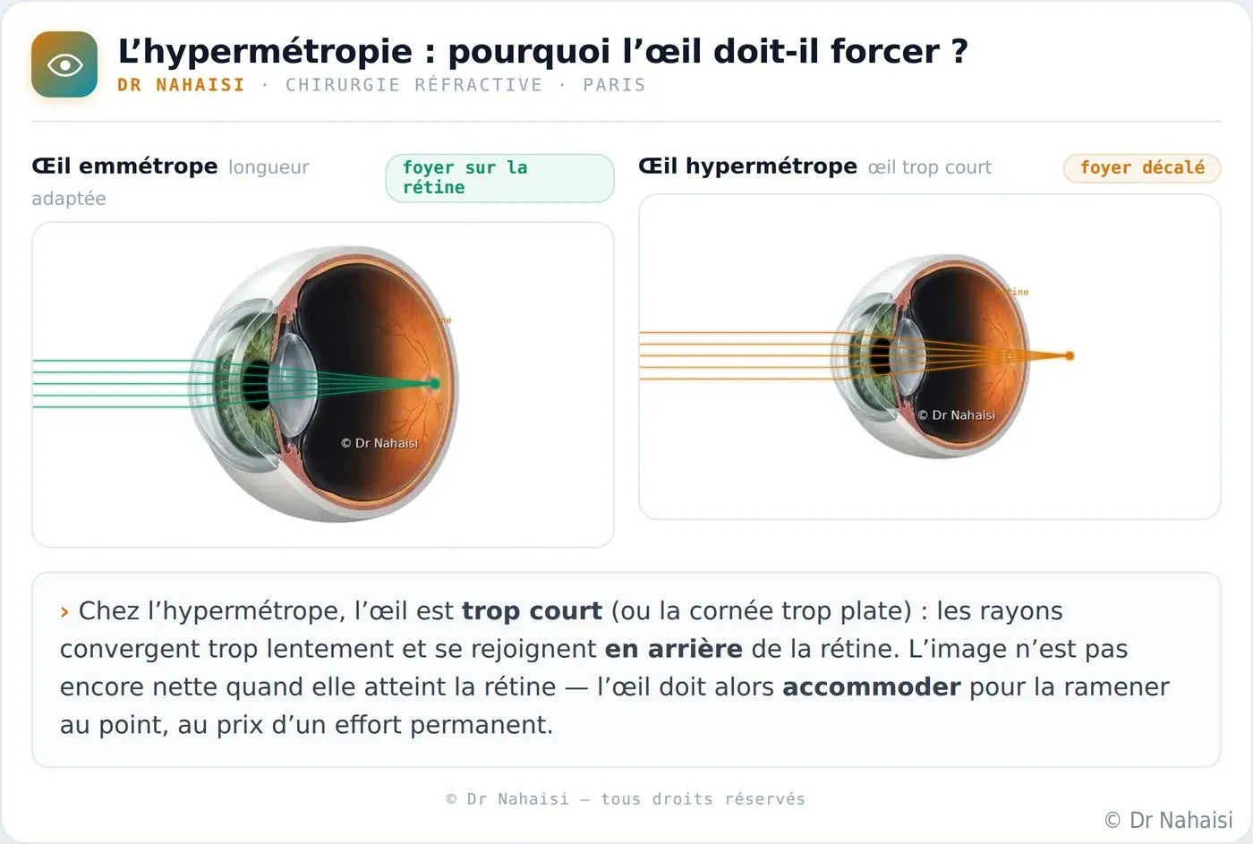
Comparison of an emmetropic eye, a nearsighted eye and a farsighted eye. In an emmetropic eye, the rays converge exactly on the retina. They converge in front of it in a nearsighted eye (too long, or cornea too steep), and behind it in a farsighted eye (too short, or cornea too flat). © Dr Nahaisi.
The length of the eye cannot be changed; it is a fixed anatomical parameter. The only variable that can be accessed surgically, and non-invasively, is the curvature of the cornea. By reshaping it, we give the light rays back the right landing point on the retina. The laser touches neither the lens, nor the retina, nor the inside of the eye.
ANATOMY OF THE CORNEA
The layers of the cornea: where does the laser act?
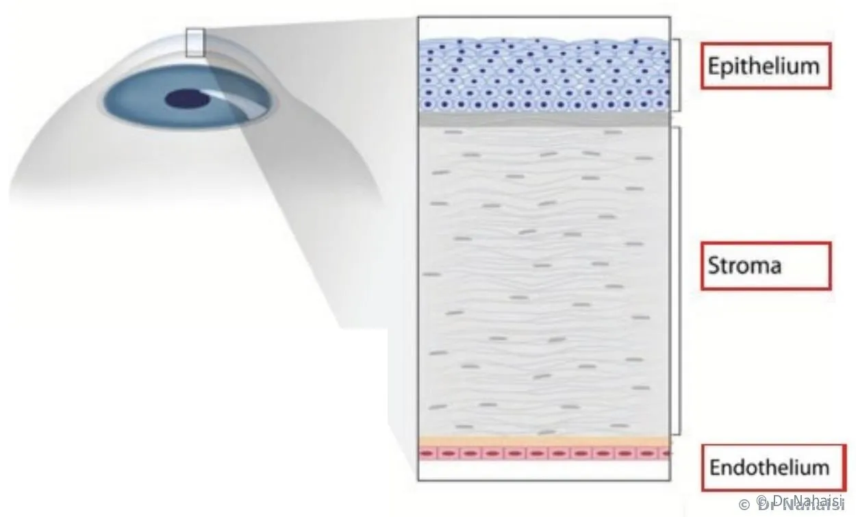
The cornea is on average 525 microns thick, or about half a millimeter. It is made of three stacked layers, separated by two membranes. Two of these layers are involved in the laser procedure. The third, the deepest, is never touched.
The epithelium, the surface layer
The epithelium is the outermost layer of the cornea, and is about 50 microns thick. It is a non-keratinized stratified squamous epithelium, with an architecture close to that of the skin of the body, but thinner and more regular. It has five to seven stacked cell layers, organized into three types. A single basal layer of cuboidal cells rests on Bowman’s membrane. Above it come two to three layers of so-called “wing” (intermediate) cells, then two to three layers of flattened surface cells.
Only the basal cells are mitotically active. They divide, and their daughter cells gradually migrate toward the surface, differentiating successively into wing cells and then surface cells, before being shed into the tear film. Complete renewal of the epithelium takes about seven days in the absence of injury.
TransPRK relies on this natural regeneration: the excimer first ablates the epithelium to expose the stroma, then sculpts the stroma. After the laser, the corneal surface closes over in 24 to 72 hours as epithelial cells migrate in from the periphery, and full stratification of the epithelium is completed over one to two weeks.
In LASIK and SMILE too, the epithelium heals, but only along the “side cut”, the peripheral cut line at the edge of the flap (LASIK) or of the micro-incision through which the lenticule is extracted (SMILE). This peripheral healing also takes a few days.
The stroma, the deep and strong part
The stroma makes up about 90% of the thickness of the cornea. It is composed of highly organized collagen fibers, which provide both the transparency and the mechanical strength of the whole. Unlike the epithelium, the stroma does not regenerate after ablation.
The laser changes the corneal curvature at the level of the stroma. Because the stroma does not heal by returning to its original shape, the new curvature is maintained over time. This is what makes refractive surgery long-lasting. All four techniques (LASIK, TransPRK, SMILE, PresbyLASIK) change the shape of the stroma; they differ only in how it is accessed.
The endothelium, the inner surface, never touched
The endothelium is the deepest layer of the cornea. It acts as a pump: it keeps the cornea dehydrated, and therefore transparent. No laser refractive surgery touches the endothelium. The laser never goes that deep into the cornea.
The two membranes separating the layers are Bowman’s membrane (between the epithelium and the stroma) and Descemet’s membrane (between the stroma and the endothelium). They act as transitions but have no direct bearing on the principle of laser treatment.
Ref.: Bourges JL et al., eds., « La cornée, Rapport de la Société Française d’Ophtalmologie », Elsevier-Masson, 2015. Wilson SE et al., « Epithelial Basement Membrane Wound Healing », Experimental Eye Research, 2017. Ehlers N et al., « Morphological evaluation of normal human corneal epithelium », Acta Ophthalmologica, 2010.
THE PRINCIPLE OF LASER CORRECTION
How laser eye surgery corrects vision: reshaping the cornea
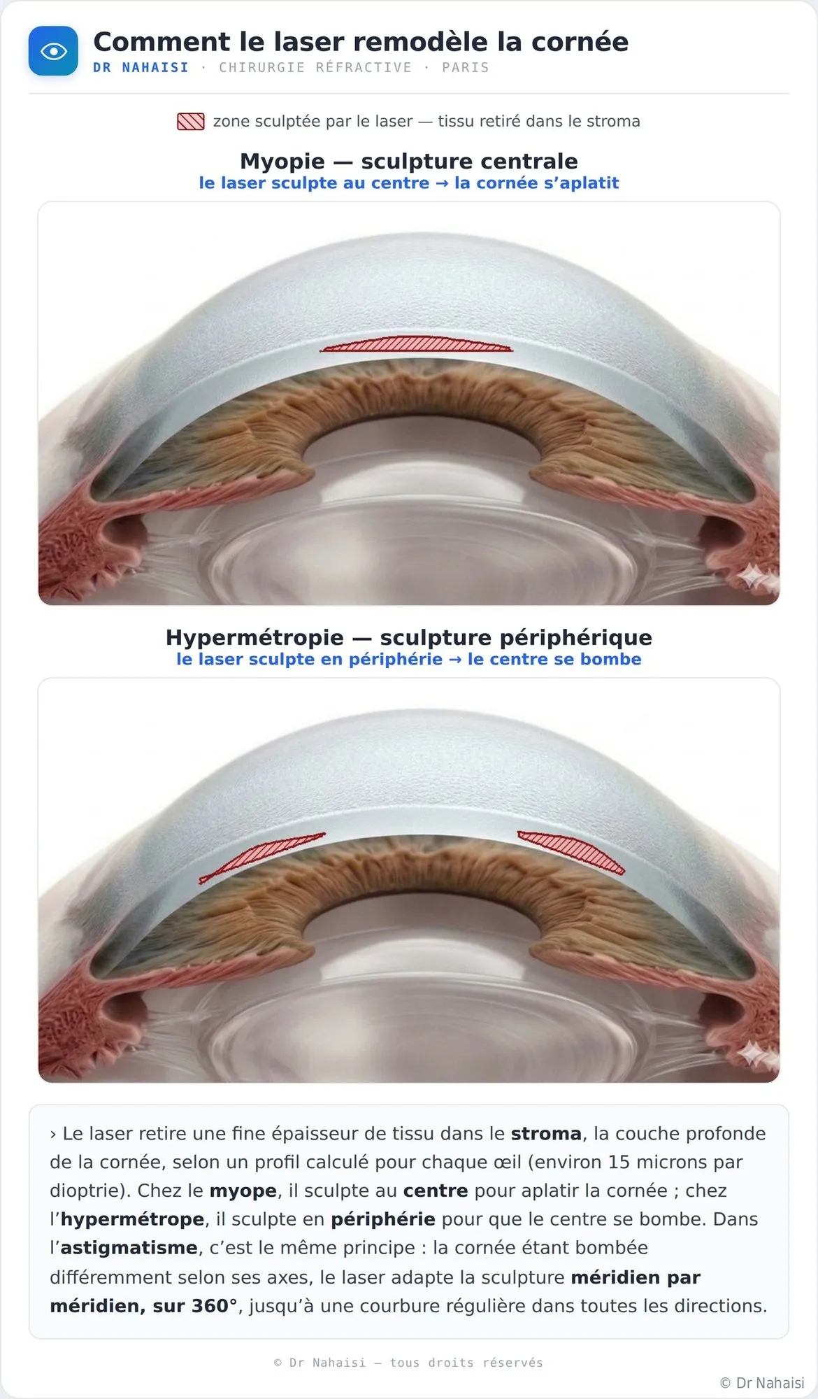
The goal is to change the shape of the cornea so that light rays converge on the retina again, without glasses or contact lenses. To do this, the laser removes a very thin layer of corneal tissue, following a profile calculated for each eye. The change sought depends on the refractive error to be corrected.
Myopia: flattening the cornea
In a nearsighted eye, the eye is too powerful, either because it is too long or because the cornea is too steep. The rays converge in front of the retina. The laser removes a small thickness from the center of the cornea to flatten it, which reduces its converging power and brings the focal point back onto the retina. Count about 15 microns of ablation per diopter of myopia treated.
Hyperopia: steepening the cornea
In a farsighted eye, the eye is not powerful enough. The rays converge behind the retina. The laser then removes tissue from the periphery of the cornea, to make it steeper in the center, which increases its converging power. The treatment is technically more demanding than for myopia, and its limits depend on the corneal curvature.
Astigmatism: evening out the curvature
In an astigmatic eye, the cornea is not evenly curved. It is steeper along one axis than along the other, a bit like a football rather than a soccer ball. The laser then sculpts an asymmetric profile, which reduces this difference in curvature between the two axes and gives the cornea a regular geometry again.
Presbyopia: creating several focusing zones
In a presbyopic eye, the lens has lost its flexibility and can no longer accommodate to see up close. The laser does not restore accommodation. But it can create zones of different curvature on the cornea (between the center and the periphery), which allow the eye to see clearly at several distances. This is the principle of PresbyLASIK.
THE TWO LASERS USED AT THE INSTITUT VOLTAIRE
Femtosecond and excimer: two lasers, two distinct roles
Two complementary lasers are used at the Institut Voltaire for refractive surgery. They do not have the same function and do not act on the same layers of the cornea.
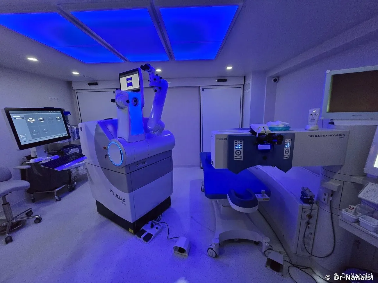
The operating room at the Institut Voltaire. On the left, the Zeiss Visumax 800 femtosecond laser (ultra-precise cuts: the LASIK flap, the SMILE lenticule); on the right, the Schwind Amaris 750S excimer laser (sculpting the stroma by photoablation).
The femtosecond laser: Visumax 800 (Zeiss)
The femtosecond is a cutting laser with micrometer precision. Its pulses last a millionth of a billionth of a second, hence its name. They create a clean tissue separation inside the cornea, without any mechanical incision. The cornea is never touched by an instrument.
The Institut Voltaire is equipped with the Visumax 800, the latest generation of Zeiss femtosecond laser. It is used in two techniques:
- In LASIK, it cuts a corneal flap, a thin surface layer about 110 microns thick, which is then lifted to expose the stroma to the excimer treatment. The cut takes 8 to 10 seconds per eye, at low pressure and painlessly.
- In SMILE, it performs the entire treatment, sculpting a lenticule inside the stroma, which is then extracted through a peripheral micro-incision.
The excimer laser: Schwind Amaris 750S
The excimer is the laser that sculpts the cornea. It emits ultraviolet light at a precise wavelength (193 nanometers), which removes corneal tissue by photoablation: the light instantly breaks the molecular bonds of the outermost layers of the tissue, which are vaporized without significant heat transfer. The surrounding corneal tissue stays cold.
The Schwind Amaris 750S is the excimer platform used at the Institut Voltaire. Its firing rate and its eye-tracking system allow a fast treatment (about 2.5 seconds per diopter of myopia treated) that stays aligned with the eye at all times, including during involuntary microsaccades of the gaze.
The excimer is used in LASIK, TransPRK and PresbyLASIK.
Summary: which laser for which technique
| Technique | Femtosecond (Visumax 800) | Excimer (Schwind Amaris 750S) |
|---|---|---|
| LASIK | Cuts the corneal flap | Sculpts the stroma |
| TransPRK | Not used | Ablates the epithelium, then sculpts the stroma |
| SMILE | Performs the entire treatment | Not used |
| PresbyLASIK | Cuts the corneal flap | Multifocal sculpting of the stroma |
Understanding aberrometry and customized treatment
CUSTOMIZED TREATMENT
Why your laser treatment is not standard: aberrometry
A glasses prescription is an average value. When we say an eye is at -3, it means its average correction is -3 diopters. But your eye is not uniformly at -3 across its entire surface. It is a complex surface, made of countless points that each have their own correction: -2.90 here, -3.10 a little further on. These variations are called optical aberrations.
The laser, however, does not treat an average value. It treats the cornea point by point, adapting to the actual aberrometric profile of each eye. The Schwind platform allows this personalized treatment, called an aberration-free profile, which calculates the exact ablation to apply at each point of the cornea.
Aberrometry is measured a first time at the pre-operative assessment, then again on the day of the procedure, just before the laser treatment. It is this last, most up-to-date measurement that is fed into the laser to program the treatment. The result is finer than a correction based on an average value, such as the one applied in glasses.
Learn more about aberrometry and customized treatment
WHAT THE LASER DOES, AND WHAT IT DOES NOT DO
Misconceptions and facts about the corneal laser
Does the refractive surgery laser burn the cornea?
No. This fear is common and understandable; the word laser immediately suggests heat. Excimer photoablation, however, is not combustion: the ultraviolet light breaks the molecular bonds of the surface layers of the tissue, which are vaporized instantly, without significant heat transfer. The cornea itself stays cold throughout the laser treatment.
You may notice a slight smell during the laser treatment, a bit like burnt dust. It comes from the reaction of the beam with the surrounding air, not from your cornea.
Can the refractive surgery laser burn the skin of the face?
No. This question comes up fairly often in consultation, from patients who have in mind the scene in Final Destination where a laser eye operation turns into a disaster and burns the patient’s face. That is a movie scenario, unrelated to the physical reality of the refractive surgery laser.
The laser beam is focused on a very precise spot on the cornea, and its energy is entirely absorbed by the first layers of tissue. It can neither escape the surgical field nor travel any distance. During the procedure, the eye is positioned under the laser by a fixation and eye-tracking system, and the rest of the face is covered by a sterile surgical drape. Your skin is never exposed to the beam. No physical mechanism would allow the laser to burn a surface located outside its focal point.
Does the laser go inside the eye?
Never. Laser refractive surgery acts only on the cornea, the outer, transparent membrane of the eye. The laser does not penetrate inside the eye, and touches neither the lens, nor the retina, nor the optic nerve.
Is the correction achieved long-lasting?
Yes. The stromal tissue removed does not regenerate, so the new shape given to the cornea is long-lasting. The corrected vision is most often maintained for decades.
There are rare cases of regression, mainly in highly farsighted patients and with older generations of lasers. A secondary enhancement can then correct them.
Does the laser slow the progression of myopia?
No. The laser reshapes the cornea to make vision clear without optical correction. It does not change the length of the eye, which, in nearsighted people, is the parameter that may still change in young adults. That is why I generally wait until your prescription is globally stable before operating, without waiting for a specific length of time. Don’t worry about micro-variations, which are natural fluctuations.
Does the eye remain intact after surgery?
Yes. No implant and no intraocular lens is placed in LASIK, TransPRK, SMILE or PresbyLASIK. The laser reshapes the cornea, and that is all. The eye keeps its internal architecture.
THE FOUR REFRACTIVE SURGERY TECHNIQUES
Which technique for which eye?
The laser principle is the same for all techniques. What changes is how the stroma is accessed in order to sculpt it, and how the eye heals afterward.
| Technique | Principle | Main indications | Visual recovery |
|---|---|---|---|
| LASIK | Corneal flap cut with the femtosecond laser, stroma sculpted with the excimer laser, flap repositioned at the end of the procedure | Myopia, hyperopia, astigmatism; cornea of sufficient thickness | Functional vision from the next day |
| TransPRK | No flap: the excimer first ablates the epithelium, then sculpts the stroma. The epithelium regenerates within a few days. | Thin corneas, profiles at risk of eye trauma (combat sports, law enforcement, military) | About one week before returning to work |
| SMILE | Lenticule sculpted with the femtosecond laser inside the stroma, then extracted through a micro-incision | Myopia, with or without low astigmatism | Functional vision from the next day |
| PresbyLASIK | LASIK with multifocal sculpting of the stroma: several zones of different curvature | Presbyopia in emmetropic or farsighted patients | Gradual adaptation over a few weeks |
No technique is better than the others in absolute terms. There is the one that suits your eye and your prescription. Your lifestyle matters too. The pre-operative assessment decides based on objective criteria.
WHAT IF LASER IS NOT POSSIBLE?
When the corneal laser reaches its limits
The laser corrects most refractive errors, but it has its limits. The usual thresholds at the Institut Voltaire are about -10 diopters for myopia (sometimes -12 depending on the cornea), +7 to +8 diopters for hyperopia, and -6 diopters for astigmatism.
Beyond these, or if the cornea is too thin or too irregular, other options exist. The phakic implant (ICL, implantable collamer lens) is a lens placed inside the eye, behind the iris, which corrects high refractive errors without touching the cornea. When this option is best suited to your case, I refer you to a specialized colleague.
My role is to identify beforehand when laser is possible, and when it is not. I would rather not operate than take a risk with your eyes.
Frequently asked questions
Your frequently asked questions about the laser and refractive surgery
Does the excimer laser used in refractive surgery burn the cornea?
No. The excimer works by photoablation: its ultraviolet light breaks the molecular bonds of the surface layers of the corneal tissue, which are vaporized instantly, without significant heat transfer. The cornea itself stays cold throughout the laser treatment. The smell of burnt dust you may notice during the procedure comes from the reaction of the beam with the surrounding air, not from the eye.
Can the refractive surgery laser burn the skin of the face during the operation?
No. The scene in Final Destination where a laser eye operation turns into a disaster and burns the patient’s face is pure fiction. The refractive surgery laser beam is focused on a precise point on the cornea, and its energy is entirely absorbed by the first microns of corneal tissue. It cannot escape the surgical field or reach the skin. The eye is held precisely under the laser during the treatment, and the face is covered with a sterile surgical drape.
Does the refractive surgery laser go inside the eye during the operation?
Never. Laser refractive surgery acts only on the cornea, the transparent membrane at the front of the eye. The lens, the vitreous, the retina and the optic nerve are not touched. No intraocular structure is exposed.
Which layer of the cornea does the laser act on in refractive surgery?
The laser sculpts the stroma, the deep and strong layer that makes up about 90% of the corneal thickness. In TransPRK, the excimer first ablates the surface epithelium, which then regenerates on its own. The endothelium, at the back of the cornea, is never touched, whatever the technique.
How many microns of cornea does the laser remove during refractive surgery?
For myopia, count about 15 microns of stromal ablation per diopter corrected. A myopia of -3 diopters therefore requires about 45 microns of stroma, on a cornea that measures 525 on average. Ablation volumes are of the same order of magnitude for hyperopia and astigmatism. That is very little, and it is why pre-operative corneal thickness is one of the central criteria for eligibility.
Does the cornea regenerate after laser surgery?
The epithelium, the surface layer of the cornea, regenerates naturally after ablation: the surface closes over in 24 to 72 hours and full stratification re-forms over one to two weeks. This is what allows TransPRK to heal, and what closes the peripheral “side cut” in LASIK and SMILE. The stroma, on the other hand, does not regenerate. The new shape given to the cornea by the laser is therefore long-lasting, and the visual correction is maintained over time.
Why does refractive surgery use two different lasers?
Because they do not have the same function. The femtosecond laser is an ultra-precise cutting laser, used to create the LASIK flap or the SMILE lenticule. The excimer laser is a sculpting laser working by photoablation, used to reshape the stroma. Cutting and sculpting require two different technologies, which complement each other in LASIK and are used alone in TransPRK (excimer) or SMILE (femtosecond).
What is the difference between the femtosecond laser and the excimer laser in refractive surgery?
The femtosecond laser cuts. Its ultra-short pulses create a clean tissue separation inside the cornea, without mechanical contact. The excimer laser, on the other hand, sculpts: its ultraviolet light vaporizes tissue by photoablation, without heat. LASIK uses both, one after the other; TransPRK uses only the excimer; SMILE uses only the femtosecond.
Which vision problems can the refractive surgery laser correct?
The laser corrects myopia, hyperopia, astigmatism and presbyopia. The usual thresholds at the Institut Voltaire are about -10 diopters for myopia (sometimes -12 depending on the cornea), +7 to +8 diopters for hyperopia, and -6 diopters for astigmatism. Beyond these limits, other surgical options exist, such as the ICL phakic implant, and a referral to a specialized colleague may be offered.
Can laser surgery be repeated several times on the same eye?
Yes. A laser enhancement is possible if the initial correction has slightly regressed, or if presbyopia or another refractive error has appeared later. It is even included in the care package for the first year. Several years later, it remains technically possible, provided enough corneal thickness remains. Everything depends on the initial treatment and the condition of your cornea at the time the question arises.
Is the result of laser eye surgery long-lasting?
The stromal tissue removed does not regenerate, so the new corneal shape is stable over time: the correction achieved is long-lasting. But the eye goes on living, and presbyopia in particular appears after age 40-45 in everyone, whether they have had surgery or not. A secondary enhancement remains possible if things change.
Does laser eye surgery hurt?
No. Whatever the technique, the surgery is performed under topical anesthesia, with a few anesthetic drops placed on the eye. During the procedure, there is no pain and no injection. The immediate aftermath differs by technique: comfortable from the moment you leave with LASIK or SMILE, a little more uncomfortable for the first 48 to 72 hours with TransPRK, while the epithelium heals.
BOOK AN APPOINTMENT
It remains to find out whether your eye is a candidate
The principle is the same for all techniques. The one that suits your eye, however, can only be decided after a complete assessment. A single consultation is enough in the vast majority of cases.
Book an appointment on Doctolib
Phone and WhatsApp: 01 84 80 28 04 · Email: nahaisi@laservisionparis.com · Doctolib messaging
Download the information booklet (PDF)
Learn more about the pre-operative assessment
Consultation locations
| Location | Address | Hours | Metro |
|---|---|---|---|
| Centre Santé Plus, Paris 10th (10e arrondissement) | 12 rue du Faubourg Saint-Martin, 75010 Paris | Thursday afternoon | Strasbourg-Saint-Denis (4, 8, 9) |
| Cabinet Ophtavie, Paris 19th (19e arrondissement) | 37 rue Archereau, 75019 Paris | Monday afternoon | Crimée (7) |
| Institut Laser Ophtalmologique Voltaire, Paris 11th (11e arrondissement) | 5 rue La Vacquerie, 75011 Paris | Wednesday afternoon | Voltaire (9), Philippe Auguste (2) |
The surgical procedure itself takes place exclusively at the Institut Voltaire (Paris 11th), on Friday mornings, sometimes on Wednesday afternoons.
Author: Dr Steven Nahaisi, ophthalmic surgeon, specialist in laser refractive surgery, Institut Laser Ophtalmologique Voltaire, Paris 11th.
Your surgeon
Dr Steven Nahaisi
Ophthalmic surgeon dedicated exclusively to laser refractive surgery. He operates at the Institut Laser Ophtalmologique Voltaire and consults in three Paris offices. Consultations in English.