Procedure
When Laser Surgery Isn't Possible: ICL Implant and Alternatives
The vast majority of patients who come to see me are candidates for laser surgery. Some are not. A cornea that is too thin, early keratoconus, a correction beyond what the laser can achieve, a retina or a lens that changes the picture. When that is the case, I say so in consultation, I explain why, and I show you what remains possible.
Dr Steven Nahaisi
Updated on 15 October 2026 · 34 min read
Because there is almost always something left. The phakic implant, also called ICL, corrects refractive errors that the laser cannot treat, without touching the cornea. Lens surgery with an implant addresses other situations, when age or early cataracts come into play. And for keratoconus, there is a whole course of care, made up of monitoring, controlling eye rubbing and sometimes additional corneal procedures, such as cross-linking or intracorneal rings.
On this page, I explain why an eye may not be a candidate for laser surgery, what the assessment can detect, how a phakic implant works, in which situations we turn to the lens, and what I do when I decide not to operate.
On this page11 sections
- 02WHY AN EYE MAY NOT BE A CANDIDATE FOR LASER SURGERY
- 03WHAT THE PRE-OPERATIVE ASSESSMENT CAN DETECT
- 04WHAT I DO WHEN I DECIDE NOT TO OPERATE
- 05THE ICL PHAKIC IMPLANT
- 06LENS SURGERY WITH AN IMPLANT
- 07KERATOCONUS: MONITOR, STABILIZE, CORRECT
- 08A NO TODAY IS NOT A NO FOREVER
- 09HOW A REFERRAL TO A COLLEAGUE WORKS
- 10THE CONSULTATION
- 11FAQ
- 12BOOK AN APPOINTMENT
WHY AN EYE MAY NOT BE A CANDIDATE FOR LASER SURGERY
Contraindications to laser refractive surgery
The laser acts on the cornea. It removes a thin layer of tissue to give it a new curvature. This approach has its own limits: there must be enough tissue, that tissue must be healthy and regular, and the defect to be corrected must fit within the available margin. Here are the situations I encounter most often.
Keratoconus, or even suspected keratoconus
This is the first contraindication, and by far the most important. Keratoconus is a progressive deformation of the cornea, which thins and bulges into a cone. Laser surgery performed on a predisposed cornea can trigger or accelerate this deformation. This is called post-operative ectasia, and it is the adverse effect every refractive surgeon seeks to avoid.
Advanced forms are easy to recognize. Early forms, those that do not yet cause any symptoms, require a trained eye: a subtle asymmetry between the two halves of the cornea, a localized thinning, an irregularity of the epithelium over a bulging area, a back surface that is slightly out of line. My hospital training in corneal surgery, with more than 350 corneal transplants performed, taught me to spot these signs and to tell real warnings from false ones.
When I see confirmed keratoconus, I do not perform laser surgery. When I see a cornea that is merely suspicious, without certainty, I prefer to wait and see the patient again rather than take the risk.
A cornea too thin for the correction needed
A cornea measures 525 microns on average. The standard threshold for LASIK is 500 microns of pre-operative thickness. Below that, TransPRK often takes over, since it does not create a flap and preserves more thickness.
Thickness alone never decides everything. A regular cornea at 490 microns may be more suitable for laser surgery than a cornea at 520 microns that shows irregularities. What matters is the combination of thickness, regularity, curvature and the correction to be removed.
An insufficient residual stromal bed
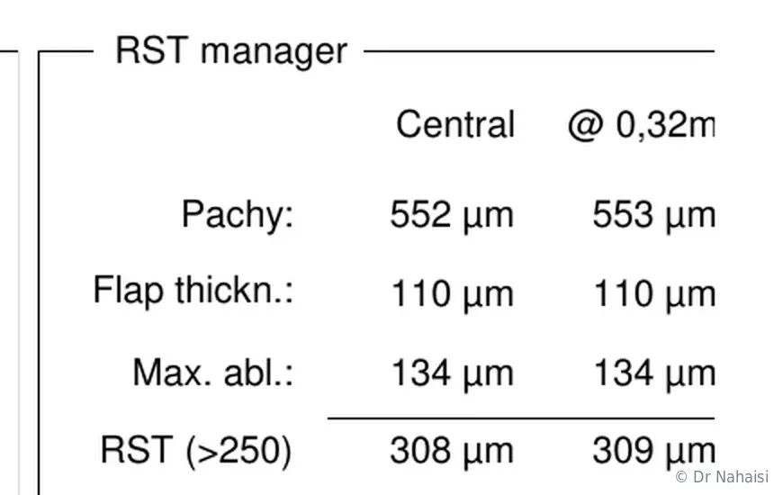
The residual stromal bed is the thickness of intact cornea that remains under the treated area. It is what guarantees the mechanical strength of the eye over decades. In LASIK, we count about 110 microns for the flap, then about 15 microns per diopter of myopia corrected. What remains underneath is the residual bed.
Standard recommendations set the limit at 280 microns. My personal limit is 300 microns, deliberately stricter. When the calculation shows that we would fall below this mark, I do not perform LASIK. Depending on the case, we switch to TransPRK, which saves the tissue of the flap, or we move away from laser surgery.
The "RST manager" of the Schwind platform, on a real anonymized case. The software starts from the measured thickness of the cornea (552 microns), subtracts the thickness of the flap (110 microns) then the ablation depth needed for the correction (134 microns), and displays what will remain of the stromal bed: 308 microns here, above the safety threshold. It is this calculation, done for each eye before the procedure, that decides candidacy for LASIK.
A correction too high for the available thickness
The higher the correction, the more tissue the laser removes. Beyond a certain level, the available margin is no longer enough. The thresholds I apply at the Institut Voltaire are around -10 diopters for myopia, sometimes up to -12 on very thick corneas, +7 to +8 diopters for hyperopia, and -6 diopters for astigmatism.
In between, there is a zone where a compromise can be chosen: treat as much as the cornea allows and accept a slight residual error. A nearsighted patient at -12 might aim for a result around -1, a farsighted patient at +7 a result around +1. The gain remains considerable compared with the thick glasses they started with. I always present this compromise clearly, with its advantages and its limits, and the decision is made together.
A corneal curvature that stands in the way
In hyperopia, the limit comes mainly from curvature. The laser has to steepen the cornea, and there is a limit beyond which we do not go, around 50 diopters of post-operative curvature. A cornea that is already steep to begin with reaches this limit sooner. In myopia, it is the opposite: a cornea that is already very flat does not tolerate being flattened further.
Severe dry eye
Moderate dry eye is not a contraindication to surgery. It guides the choice of technique and the post-operative treatment. Severe dry eye, on the other hand, with a truly damaged ocular surface, must be treated first. Preservative-free artificial tears, tear gels in the evening, sometimes doxycycline tablets when there is an associated dermatological condition. We then reassess. The situation may become compatible with surgery again, often surface surgery rather than LASIK.
A retinal or lens condition
The laser corrects the cornea. It changes nothing about what lies behind it. A progressive retinal condition, unstabilized diabetic retinopathy, a peripheral lesion that must be treated first: these require dealing with the problem in the right order. That is why the dilated fundus exam is part of the assessment.
The lens matters just as much. From around age fifty, it begins to lose its transparency in some patients. If early cataracts are present, correcting the cornea with the laser no longer makes much sense: the visual discomfort will come from the lens, and it will have to be operated on one day anyway. Better to treat the right part of the eye directly.
A prescription that has not stabilized
We operate on a stable prescription. In a young patient whose myopia has changed over the last two years, I prefer to wait, repeat the measurements in six months or a year, and decide afterward.
I do qualify this notion of stability, though, because it is often misunderstood. Perfect stability does not really exist. The eye accommodates, meaning it can strain to see up close and create temporary myopia, sometimes even in distance vision. Five exams on the same day can show differences of half a diopter in one direction or the other. What I am looking for is a clear, documented trend, not a fixed number.
The general medical context
Some situations call for a careful, case-by-case assessment, without being absolute contraindications. A progressive autoimmune disease affecting the ocular surface, a history of corneal herpes with a risk of reactivation, poorly controlled diabetes, ongoing immunosuppressive treatment, advanced glaucoma. I look at each file on its own merits, and I discuss it with the doctor who follows you when that is useful.
Pregnancy is a special case. Hormonal fluctuations can temporarily change the refraction, and I do not operate during this period, as a precaution. Breastfeeding, on the other hand, is not a problem. And you don't have to wait until you have had all your children to have surgery.
WHAT THE PRE-OPERATIVE ASSESSMENT CAN DETECT
The assessment is what decides, not the prescription on your glasses
None of these contraindications can be seen with the naked eye, and none can be guessed from your glasses prescription. They are detected with tests, all performed during the pre-operative consultation, which lasts about an hour.
Here is what each test contributes to the decision.
| Test | What it can detect |
|---|---|
| Corneal topography (front and back surfaces) | Early keratoconus, asymmetry, irregularity, acquired deformation |
| Pachymetry | Total corneal thickness, margin available for treatment |
| Epithelial mapping | Localized thinning of the epithelium, an early sign of fragility |
| Calculation of the residual stromal bed | Feasibility of LASIK, switch to TransPRK, or moving away from laser surgery |
| Keratometry | Limits of steepening or flattening of the cornea |
| Aberrometry | Optical quality of the eye, programming of the customized treatment |
| Ocular surface exam | Dry eye, allergic tendency, blepharitis |
| Dilated fundus exam and retinal photography | Retinal disease, peripheral lesions in highly nearsighted patients |
| Lens exam | Early cataracts, transparency of the lens |
| Intraocular pressure | Ocular hypertension, early glaucoma |
| Questions about eye rubbing | Behavior that puts the cornea at risk of deformation |
I often say in consultation that I am not trying to confirm that a cornea is operable. I am looking for the slightest reason why it might not be. Refractive surgery is elective surgery, and my duty is to make it as safe as possible.
The pre-operative assessment: everything you need to know
Ref.: Randleman JB. et al., "Risk assessment for ectasia after corneal refractive surgery," Ophthalmology, 2008. Ambrósio R. Jr. et al., "Integration of Scheimpflug-based corneal tomography and biomechanical assessments," J Refract Surg, 2017.
WHAT I DO WHEN I DECIDE NOT TO OPERATE
A refusal that is explained, reasoned, and followed by a proposal
Sometimes I say no. I say it clearly, directly and without leaving any doubt, because a patient who leaves with a vague answer will simply go looking for a yes elsewhere.
I would rather not operate than take the slightest risk with your eyes.
I show you the test results
When the answer is no, I put the images in front of you. The topography, the thickness map, the calculation of the residual bed. I show you the exact point that stands in the way and I explain what would happen if we ignored it. This transparency has a practical purpose: it gives you what you need to understand, and to recognize a different message if you consult elsewhere.
Borderline cases are discussed as a team
At the Institut Laser Ophtalmologique Voltaire, I work with other surgeons specializing in refractive surgery. Every week, we discuss together the files that raise questions.
This three-way consultation changes the nature of the decision. When we unanimously confirm that you are a candidate, you can move forward with confidence. When we decide to wait, it is because caution requires it, and it is not the isolated opinion of a single surgeon.
I suggest a next step
A refusal without a proposal is of little use. Depending on the reason for the refusal, the next step takes a different form. A phakic implant when the correction exceeds the limits of the laser or when the cornea cannot be touched. Lens surgery when age or cataracts call for it. Close monitoring and work on eye rubbing when the cornea shows signs of fragility. A simple postponement when the prescription is not yet stable.
In every case, you leave with an explanation, a direction, and the option to write to me again whenever you want.
THE ICL PHAKIC IMPLANT
The ICL: a lens placed inside the eye, without touching the cornea
The phakic implant is the main alternative to laser surgery. It is commonly called ICL, for Implantable Collamer Lens, after the soft, biocompatible material it is made of.
How it works
The ICL is a thin, soft lens placed inside the eye, in front of the natural lens and behind the iris. It sits in a space that already exists, without removing anything. The natural lens stays in place and continues to work normally, which explains the term "phakic," which means precisely that the eye keeps its natural lens.
The cornea is not touched. No tissue is removed, no curvature is changed, no thickness is used up. This is what makes the ICL possible where laser surgery no longer is.
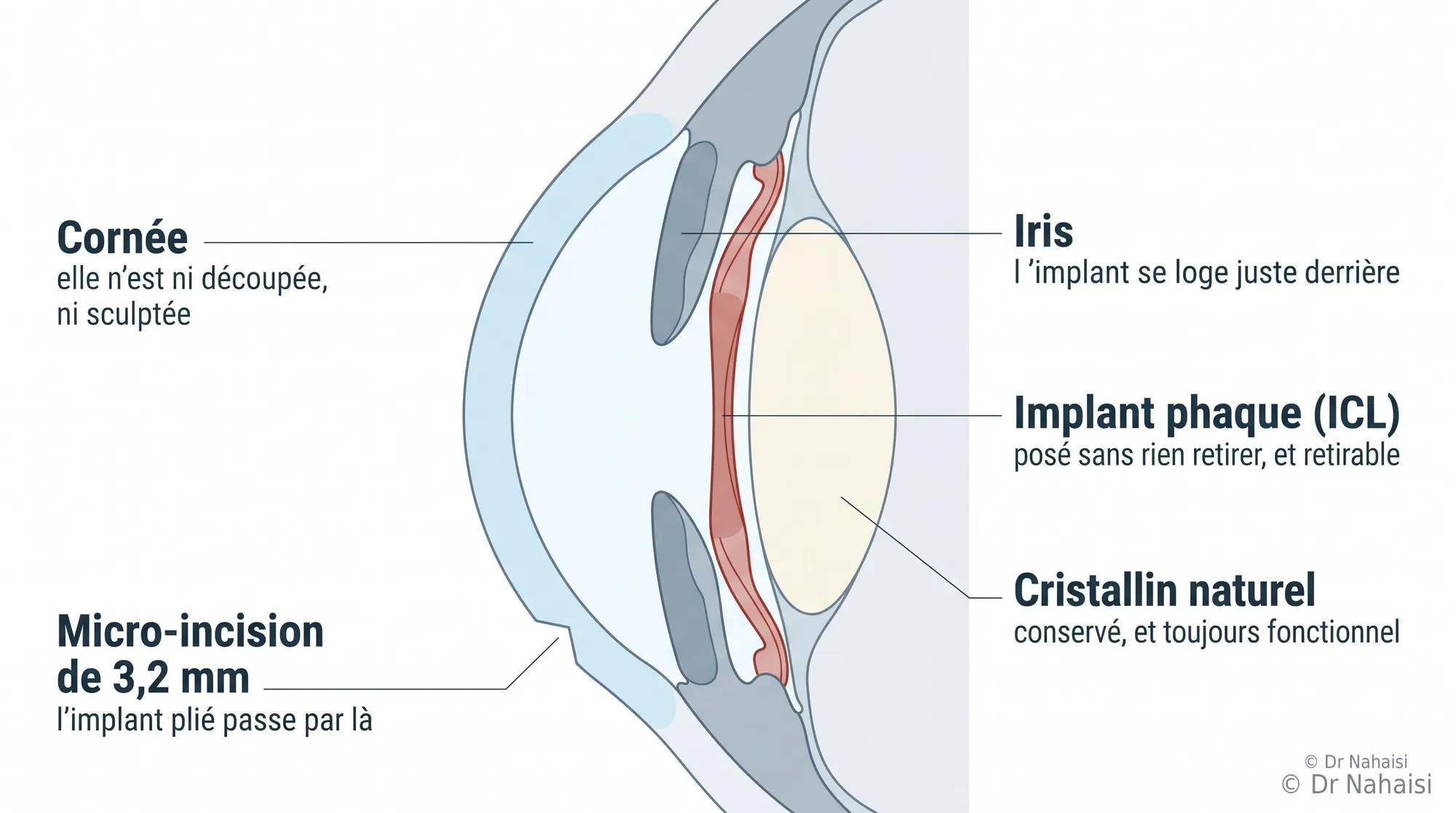
The position of the phakic implant in the eye. The cornea is neither cut nor sculpted; it stays as it is. The implant is slid in through a 3.2 millimeter micro-incision, then placed behind the iris, in front of the natural lens, which is preserved and continues to work. Nothing is removed, and the implant can be taken out if necessary.
Who it is for
The ICL is a good answer in four main situations.
High myopia. Beyond about -10 diopters, the laser reaches the limit of what the cornea can give. The ICL, by contrast, corrects much higher myopia without using up corneal tissue. This is the most common indication.
Corneas that are too thin. When the thickness or the calculated residual stromal bed allows neither LASIK nor TransPRK, the ICL gets around the problem by working inside the eye.
Corneas we prefer not to touch. A slightly irregular cornea, a worrying profile, a history that makes corneal surgery undesirable.
Corrections the laser cannot reach. Some high astigmatism combined with high myopia falls into this category. There are toric implants, which correct myopia and astigmatism at the same time.
The ICL also corrects hyperopia, but with an important caveat that I discuss below.
A point of method: when the cornea is suitable for laser surgery, I prefer the laser. Surgery that stays outside the eye is always preferable to surgery that goes inside it, for a comparable result. The ICL comes into its own when the laser is no longer the right option, not as a convenience substitute.
How the procedure goes
The procedure is done in the operating room, under general anesthesia. An anesthesia consultation is scheduled beforehand, which sets it apart from corneal laser surgery, where it is not needed.
The surgeon makes a 3.2 millimeter micro-incision at the edge of the cornea. The folded implant is injected through this incision, then unfolds on its own inside the eye. It is then positioned behind the iris, in its natural place. The incision is so small that it closes on its own, with no stitches.
The procedure takes a few minutes per eye. Both eyes are not always operated on the same day; it depends on the surgeon's habits and on the case.
Visual recovery is fast, often within the first few days. Post-operative treatment with eye drops lasts about a month. After that, life returns to normal, sports included, diving included. The only recommendation that remains actually applies to everyone, whether they have had surgery or not: avoid violent blows to the eye.
Quality of vision
This is one of the strengths of the ICL, and I speak about it frankly with patients who are hesitating between a partial-correction laser treatment and an implant. The implant causes neither haze, the slight cloudiness that can follow surface surgery for a high correction, nor regression related to healing. The optical quality achieved on high myopia is often excellent. And if the myopia were to change later, an additional laser correction would remain possible.
On the other side, there is a small intraocular risk, which I detail below. No option is perfect, and the choice is discussed case by case.
Reversibility
The implant is designed to stay in place for life, or at least until a cataract develops. It can also be removed, which is not the case with a laser treatment. This reversibility reassures many patients, and it is real, even though removal remains a rare possibility, reserved for situations where the implant causes a problem.
The day a cataract develops, the implant will be removed during lens surgery, which will then take over both functions.
Follow-up
An annual check-up is recommended after a phakic implant. It checks the intraocular pressure, the position of the implant, the condition of the natural lens and that of the retina. It is light follow-up, but it should not be abandoned.
In highly nearsighted patients, this retinal follow-up is important for another reason. Correcting vision does not change the structure of the eye. The retina of a highly nearsighted eye remains thinner and more stretched after the operation, and it deserves a regular dilated fundus exam.
The limits of the ICL
The depth of the anterior chamber. There has to be room inside the eye to fit the implant without it coming into contact with the natural lens or interfering with the circulation of the aqueous humor. This space is measured precisely before the procedure. When it is insufficient, the implant is not placed.
This explains why the ICL is offered more often to nearsighted patients than to farsighted ones. A highly nearsighted patient has an elongated eye, where there is generally enough space. A farsighted patient by definition has a shorter eye, with an anterior chamber that is often narrower. So a highly farsighted patient may be a candidate for neither the laser nor the phakic implant. This is rare, and transparency requires saying so. The only option then becomes lens surgery, generally considered from around age fifty.
Endothelial cell density. The endothelium is the innermost layer of the cornea. Its cells do not renew, and their number naturally decreases over a lifetime. Any intraocular surgery uses up a few of them. So these cells are counted before the procedure, and we do not implant an eye whose endothelial reserve is already low.
Age. The ICL is intended rather for younger people, whose natural lens is perfectly transparent. Past a certain age, when the lens begins to cloud or presbyopia has clearly set in, lens surgery becomes more logical. Placing a phakic implant only to remove the lens a few years later makes little sense.
Specific risks. Ocular hypertension can occur, most often controllable with treatment. A cataract can appear earlier. As with any intraocular surgery, there is a very low but real risk of infection. These complications remain minimal when the pre-operative evaluation is rigorous, and anticipating them is precisely the role of the assessment.
The fee
The fee for phakic implant surgery depends on the surgeon, the price of the implants and the clinic's technical facilities. I do not list it here, because it does not depend on me and an approximate figure would not serve you well. It is given to you by the colleague who takes on your case, in the form of a detailed quote that you can send to your complementary health insurance.
LENS SURGERY WITH AN IMPLANT
When the solution lies in the lens, not the cornea
The lens is the eye's internal lens, the one that allows focusing. It stiffens over the years, which causes presbyopia around age 42-45, then it gradually loses its transparency, which causes cataracts, generally after age 60-65.
When this lens becomes the main problem, there is no reason to work on the cornea. It is replaced with an implant, and vision is corrected at the same time.
In which situations
Cataracts, even early ones. If the assessment shows a lens that is beginning to cloud significantly, a corneal laser treatment would quickly lose its value: the visual discomfort would keep progressing on its own. Lens surgery then treats the cause, and the chosen implant corrects myopia, hyperopia and astigmatism along the way.
Age, in presbyopic patients. Past 55 or 60, when presbyopia has set in and a cataract is on the horizon, operating on the lens makes it possible to deal with both issues at once. This procedure is done only once in a lifetime, since the replaced lens never clouds again.
Extreme corrections with no other solution. This is the case for highly farsighted patients whose anterior chamber is too narrow for a phakic implant. Lens surgery, then called clear lens surgery, becomes the only possible route.
The implants available
The choice of implant matters as much as the surgical procedure. It is discussed with the operating surgeon, based on lifestyle and expectations.
| Type of implant | What it offers |
|---|---|
| Monofocal | A single sharp distance, generally distance vision. Reading glasses needed afterward. |
| Toric monofocal | Same principle, with correction of astigmatism. |
| Extended depth of focus | A continuous range of vision from distance to intermediate, with good comfort and few light effects. Often an excellent compromise. |
| Multifocal | Several focal points, distance and near vision without glasses in most situations, with halos more frequent in the first few months. |
There are also toric versions of most of these implants, to treat associated astigmatism.
How it goes
The procedure is done in the operating room, under local anesthesia, after an anesthesia consultation. The surgeon removes the clouded lens through a micro-incision and puts the implant in its place. Visual recovery is fast and post-operative treatment lasts about a month.
Here again, the fee depends on the surgeon, the type of implant chosen and the clinic. The cataract-related portion is covered by French national health insurance, while so-called premium implants and correction of astigmatism leave a variable out-of-pocket cost. The quote is given to you before the procedure.
Lens surgery is part of what I have learned and practiced, and I am happy to answer your questions about it in consultation. I no longer perform it routinely: I devote all my time to laser surgery. When a case calls for lens surgery, I send your test results and my analysis to a colleague for whom it is the core of their practice, and I stay in the loop.
KERATOCONUS: MONITOR, STABILIZE, CORRECT
What we do when the assessment uncovers keratoconus
Discovering keratoconus when you came to ask for laser surgery is hard news to hear. Many patients learn about it exactly this way, during a refractive surgery assessment. It is also an opportunity: the earlier the diagnosis, the simpler the care.
I always separate things into two almost independent parts: controlling progression on one side, correcting vision on the other. Confusing the two is the source of most misunderstandings.
Controlling progression: eye rubbing first
The most important element of all keratoconus care is stopping eye rubbing. Chronic rubbing is one of the main causes of corneal deformation.
I systematically look for the habits involved, and they are not always conscious. Rubbing in the morning on waking. Itching due to an allergy, which triggers the gesture several times a day. Sleeping on your stomach, with your head pressed into the pillow. A hand or elbow pressed under the eye during the night. Once these behaviors are identified and corrected, it is very rare for keratoconus to keep progressing.
When an allergy keeps the itching going, we treat it. That is often what makes it possible to really stop rubbing your eyes, rather than trying to resist by willpower alone.
Topographic monitoring
Follow-up relies on repeated, comparable topographies. One technical point matters: it is better to repeat the tests on the same machine, otherwise measurement differences between devices make the comparison unreliable. I have seen many false progressions that were nothing more than machine variations.
The usual schedule is a check-up every six months at first, then less often if the situation remains stable.
Cross-linking
Cross-linking involves making the cornea react with riboflavin and UV light to create additional bonds between its fibers, and thus stiffen it. The goal is to stop progression, not to improve vision.
It is a much-debated technique. Some are against it, others perform it on almost everyone. In practice, I mainly reserve it for young patients, under 25, because that is when keratoconus progresses fastest and the benefit is clearest. This is even more true for a teenager who will not really follow the instructions about rubbing. Past that age, for a stable form and in someone who has understood the mechanism and has truly stopped rubbing their eyes, I am more reserved and prefer to monitor. And if progression is demonstrated by reliable topographic comparisons despite stopping rubbing, the indication becomes legitimate at any age.
When cross-linking is indicated, I refer you to a colleague or a hospital department that performs it regularly, and I see you again afterward. Be aware that the cornea keeps changing for several months after cross-linking, which means waiting at least four to six months before talking about correction again.
Correcting vision with keratoconus
Correction is discussed separately, and several options exist side by side.
Glasses, when the deformation remains moderate and corrected acuity is good.
Rigid contact lenses, which remain the best optical solution for many cases of keratoconus. The rigid lens recreates a regular surface in front of the deformed cornea, and the visual gain compared with glasses is often significant. It is frequently the first option I suggest.
Intracorneal rings, small segments inserted into the thickness of the cornea to partially regularize it. This surgery is performed by colleagues specializing in keratoconus, to whom I refer you when the indication is confirmed.
Topography-guided laser, in selected cases. I have treated patients with mild keratoconus with the laser, with very good results. It can only be considered for stable, mildly deformed forms, often after prior cross-linking. The treatment profile is custom-calculated: I send the test results to the Schwind laboratory, which runs a simulation, and it is this response that tells whether the treatment is worth undertaking. This process takes a few weeks and sometimes requires additional measurements at the Institut Voltaire.
Doing nothing, finally, which is a real option. If your vision with both eyes suits you in daily life and the more affected eye does not really bother you, doing nothing is a reasonable choice, provided monitoring continues.
A NO TODAY IS NOT A NO FOREVER
Some negative answers have an expiration date
Not all contraindications are equal. Some are structural and will not change. Others are tied to a particular time in your life or to a parameter that can be changed.
A prescription that is not yet stable
This is the most common case in patients aged 18 to 24. The myopia has changed recently, and I believe it is better to wait. We reassess six months or a year later, with comparable measurements, and the answer often becomes yes. Waiting a little takes nothing away from the final result.
A cornea weakened by rubbing
When the topography shows an irregularity related to chronic rubbing, truly stopping rubbing your eyes may be enough to stabilize the situation. After a few months of follow-up and topographies that no longer change, surgery may become possible again, often TransPRK rather than LASIK. Age also plays a role: the cornea stiffens over the years, and I more readily agree to operate on a borderline cornea at 32 than at 20.
An ocular surface to treat first
Significant dry eye, blepharitis, poorly controlled allergy are conditions that can be treated. We treat, we wait, we reassess. Many patients who leave with a surface treatment come back a few months later in much better condition.
An ongoing pregnancy
The postponement is temporary, and it is mainly a matter of precaution. Some patients have had surgery without knowing they were pregnant, and the published series on these cases show no additional complications. The data remain limited, so I prefer to stick to this simple rule: we postpone by a few months, and nothing is lost.
What, on the other hand, will not change
Confirmed keratoconus will not become a normal cornea again. A structurally too-thin cornea will not thicken. In these situations, waiting brings nothing, and it is the alternatives you need to look at.
I always tell you which category your case falls into. A "come back in a year" and a "this won't be possible with the laser" are two different answers, and you will get my most honest opinion possible.
A word about gray areas
Not every case falls clearly on one side or the other. Some have borderline criteria: a slightly thin but perfectly regular cornea, an asymmetric topography with no other sign, a history of rubbing that stopped two years ago. Here, there is no threshold that decides for you.
What decides is the sum of everything: monitoring over time, the opinion of my colleagues at the Institut when I want a second look, your age, and what surgery truly means to you. We discuss it, and the decision is made case by case.
It is also in these cases that two surgeons may not say the same thing. When that happens, I tell you, and I explain what opinions you might hear elsewhere and what they are based on. At least you will know that the disagreement is about a judgment call, not about a fact.
HOW A REFERRAL TO A COLLEAGUE WORKS
How I pass a case on to a colleague
Intraocular surgeries, whether phakic implant or lens surgery, take place in a different setting from corneal laser surgery: a conventional operating room, an anesthesia consultation, ordering implants from a manufacturer, handling insurance coverage. Given the demand for laser refractive surgery, I chose to devote myself to it entirely, and for these procedures I rely on my colleagues at the Institut Voltaire, for whom it is the core of their practice.
Referring you to someone does not mean leaving you to fend for yourself.
What I pass on
The colleague receives your complete file, with the tests performed at the Institut Voltaire and my analysis: what I found, why laser surgery is not the right option, and what seems suited to your case. You don't have to explain everything again from the beginning, and there is no reason to repeat all the tests. That is the advantage of working at the Institut Voltaire: you often come back a few days later to the same place, to see another surgeon who already has your whole file in our database.
What you keep
You keep my contact details. You can write to me again through Doctolib messaging or by email with a question about what you have been offered, about a quote, or about a worry before the procedure. I personally answer my patients.
Many patients write to me for a second opinion before committing to surgery proposed elsewhere. I am happy to do it, and I say so when I agree with what has been proposed, which is most often the case.
What I continue to follow
I remain reachable about your case. You can write to me again for an opinion before or after the procedure, and I will answer. The follow-up itself is provided by the surgeon who operates on you; it is simpler and safer for you.
THE CONSULTATION
Come in for a consultation to find out where you stand
The only way to know whether your eyes are suitable for surgery, and with which technique, is to have the assessment. This also applies to those who have already been told no elsewhere, or who think they are not candidates without ever having had a complete exam.
The fee
The consultation is charged at the standard rate set by French national health insurance. What changes depending on the location is any fee supplement and whether you pay upfront.
| Location | Paid upfront | Details |
|---|---|---|
| Paris 10th, Centre Santé Plus | €0 | Full direct billing with your carte Vitale and complementary health insurance. You pay nothing upfront. |
| Paris 19th, Cabinet Ophtavie | €0 | Full direct billing with your carte Vitale and complementary health insurance. You pay nothing upfront. |
| Institut Voltaire, Paris 11th | €50 | €50 paid upfront, including a €30 fee supplement (above the standard rate). Your complementary health insurance reimburses at least €20, and often the full amount depending on your plan. |
No referral letter needed
You need neither a letter from an ophthalmologist nor a prescription to book an appointment. You book directly on Doctolib or by phone.
If you already have test results, topographies in particular, bring them. They are useful for comparison over time, especially when monitoring keratoconus.
And the fees for the alternatives
I do not give a fee for the phakic implant or for lens surgery, because they do not depend on me. They vary depending on the surgeon, the type of implant and the clinic's technical facilities. A detailed quote is given to you before any procedure, to send to your complementary health insurance.
The fees for my laser procedures, on the other hand, are fixed and published: €3,200 for LASIK, €2,800 for TransPRK, €3,800 for SMILE, €1,800 per eye for PresbyLASIK, i.e. €3,600 for both eyes.
Frequently asked questions
Your questions when laser surgery isn't possible
What happens if I'm not a candidate for laser eye surgery?
I explain precisely what stands in the way, with test results to back it up, and I present the alternatives. For corrections beyond the limits of the laser or for corneas that cannot be touched, the ICL phakic implant is often the right answer. When age or early cataracts come into play, it is lens surgery. For a cornea that is becoming weaker, the situation can sometimes be stabilized and the question reexamined later. In every case, I would rather not operate than take the slightest risk with your eyes.
What is an ICL phakic implant?
It is a thin, soft lens placed inside the eye, in front of the natural lens and behind the iris. It corrects myopia, hyperopia and astigmatism without touching the cornea and without removing tissue. The natural lens stays in place and continues to work. The procedure is done in the operating room, under general anesthesia, through a micro-incision a few millimeters long. It is the main alternative to laser surgery for high corrections and thin corneas.
At what level of correction is an implant needed rather than a laser?
Beyond about -10 diopters of myopia, the laser reaches the limits of what the cornea can give. On a very thick cornea, we can sometimes go up to -12. Beyond that, the phakic implant becomes the best option. The number is only a guide, however: the real decision is made by combining the correction with corneal thickness, the regularity of the cornea and the calculated residual stromal bed.
Is the ICL implant reversible?
Yes. The implant is designed to stay in place for life, or at least until a cataract develops, but it can be removed if necessary. This is a real difference from the laser, which changes the cornea permanently. Removal remains a rare possibility, reserved for situations where the implant causes a problem, ocular hypertension that cannot be controlled, for example.
Can the ICL implant be seen or felt in the eye?
No. The implant is transparent and placed behind the iris, so it is invisible from the outside. It cannot be felt either: no nerve senses it once the post-operative period is over. Patients with an implant forget it is there.
Can you play sports with a phakic implant?
Yes. After about a month of recovery, all activities resume, including diving. Water pressure poses no particular problem. The only recommendation that remains applies to everyone, whether they have had surgery or not: avoid violent blows directly to the eye.
How much does ICL implant surgery cost in Paris?
The fee depends on the surgeon, the price of the implants and the technical facilities of the clinic where the procedure is done. I do not list it here because it does not depend on me and an approximate ballpark figure would not serve you well. The colleague who takes on your case gives you a detailed quote, which you can send to your complementary health insurance to find out your coverage.
I have keratoconus. Can I still have surgery to correct my vision?
Not LASIK, in any case: laser surgery with a flap can worsen the deformation. That does not mean there is nothing to be done. Care works on two independent levels. Controlling progression relies above all on stopping eye rubbing, sometimes on cross-linking. Correcting vision relies on glasses, rigid contact lenses, sometimes intracorneal rings, and in selected cases of mild, stable keratoconus, on a custom topography-guided laser treatment. We review it together with recent topographies.
Is cross-linking systematic for keratoconus?
No, and it is a technique debated among specialists. Some apply it to almost all patients, others much more rarely. I mainly reserve it for young patients, under 25, because that is the age at which keratoconus progresses the most, and even more so for a teenager who will not follow the instructions about rubbing. In an adult who has understood the mechanism and really stops rubbing their eyes, I am more reserved: it is rare for keratoconus to progress when all rubbing factors disappear. If progression is nevertheless demonstrated by comparable topographies, the indication becomes legitimate.
Can eye rubbing really damage a cornea?
Yes. Chronic rubbing is one of the main causes of corneal deformation and acquired keratoconus. The gesture is not always conscious: rubbing on waking, allergic itching, sleeping on your stomach with your head in the pillow, a hand or elbow pressed under the eye at night. Identifying these habits and changing them is sometimes enough to stabilize a fragile cornea, and to make surgery possible after a few months of follow-up.
My cornea is too thin for LASIK. Does that mean I can't do anything?
Not at all. The standard threshold of 500 microns applies to LASIK, which uses up thickness to create a flap. TransPRK, which does not create one, makes it possible to operate on thinner corneas. That is even its most common indication. When neither is possible, the phakic implant takes over. What matters is never thickness alone, but the combination of thickness, regularity and the correction to be treated.
I have early cataracts. Is laser refractive surgery still possible?
If the lens is still transparent and the cornea healthy, laser surgery remains a good option. If the assessment shows significant early cataracts, a corneal treatment loses its value, since the visual discomfort will continue to come from the lens. We then consider lens surgery with an implant, which corrects vision and treats the cataract at the same time. This procedure is performed by specialized colleagues, to whom I pass on the file.
I was told no for laser surgery elsewhere. Is it worth getting a second opinion?
Yes, it happens regularly and it is a consultation I am happy to do. Candidacy criteria are not identical from one surgeon to another, and above all not all centers have the same techniques: a cornea turned down for LASIK may be perfectly suitable for TransPRK. Sometimes I also confirm the refusal, and I then say so with the same arguments. Bring your previous test results if you have any.
Can high myopia of -12 diopters be operated on?
It depends on your cornea. On a thick, regular cornea, laser treatment is sometimes possible up to this level, possibly with a compromise: we treat as much as the cornea allows, accepting a slight residual error on the order of -1 diopter. The gain remains considerable compared with the starting point. When the cornea does not allow it, the phakic implant is the solution, and it gives excellent quality of vision for these high corrections.
Is it better to have partial-correction laser eye surgery or an implant?
It is a real question, and I put it just like that to the patients concerned. Surface laser surgery remains possible with partial correction if the cornea is regular, but it exposes you to a residual error, to possible regression with high corrections, and it will not leave much margin for a retreatment. The implant gives superior optical quality for these corrections, with no haze or regression, with the possibility of an additional laser correction later. On the other side, it carries a small intraocular risk. There is no single answer, and the choice is yours once you have all the information.
My hyperopia is too high for the laser. Is an implant possible?
Not always, and that needs to be said frankly. The phakic implant needs enough space inside the eye to be placed. A highly nearsighted patient has an elongated eye where this space generally exists. A farsighted patient by definition has a shorter eye, with an anterior chamber that is often narrower. So a highly farsighted patient may be a candidate for neither the laser nor the phakic implant. In that case, the only possible route is lens surgery, generally considered from around age fifty.
Do I need a letter or a prescription to consult about an ICL implant or an alternative to laser surgery?
No. No letter from another ophthalmologist needed, no referral needed. You book an appointment directly on Doctolib or by phone. The consultation is charged at the standard rate set by French national health insurance, with no fee supplement, and nothing to pay upfront (direct billing) at the offices in Paris 10th and 19th. At the Institut Voltaire (Paris 11th), a €30 fee supplement applies.
If you don't perform laser surgery on me, who will follow my case?
I will, if you wish. When I refer a patient to a colleague for intraocular surgery, I pass on the complete file with my analysis, and I remain available to give my opinion both before and after the procedure. You can write to me again at any time through Doctolib messaging or by email.
BOOK AN APPOINTMENT
Find out what is possible for your eyes
The first step is the same for everyone: a consultation with a complete assessment, to find out where you stand. You leave with a clear answer: a candidate or not, with which technique, with what expected result. There is no commitment and no pressure.
I see patients at three locations in Paris:
- Institut Laser Ophtalmologique Voltaire, 5 rue La Vacquerie, 75011 Paris (Wednesday afternoons). Metro Voltaire (9), Philippe Auguste (2).
- Cabinet Ophtavie, 37 rue Archereau, 75019 Paris (Monday afternoons). Metro Crimée (7).
- Centre Santé Plus, 12 rue du Faubourg Saint-Martin, 75010 Paris (Thursday afternoons). Metro Strasbourg-Saint-Denis (4, 8, 9).
Laser surgery takes place on Friday mornings at the Institut Voltaire, sometimes on Wednesday afternoons.
Book an appointment on Doctolib
Phone and WhatsApp:01 84 80 28 04 · Email:nahaisi@laservisionparis.com · Doctolib messaging
Download the information booklet (PDF)
"I don't operate on eyes, I operate on patients."
Dr Steven Nahaisi, ophthalmic surgeon in Paris, specialist in LASIK and refractive surgery.
Author: Dr Steven Nahaisi, ophthalmic surgeon, specialist in laser refractive surgery, Institut Laser Ophtalmologique Voltaire, Paris 11th.
Your surgeon
Dr Steven Nahaisi
Ophthalmic surgeon dedicated exclusively to laser refractive surgery. He operates at the Institut Laser Ophtalmologique Voltaire and consults in three Paris offices. Consultations in English.