
Full transparency
Risks and Complications of Laser Refractive Surgery
This page brings together what I say during the consultation when we discuss risks: what is part of healing, what is a real complication, what I look at before operating, and what I do when something does not go as planned.
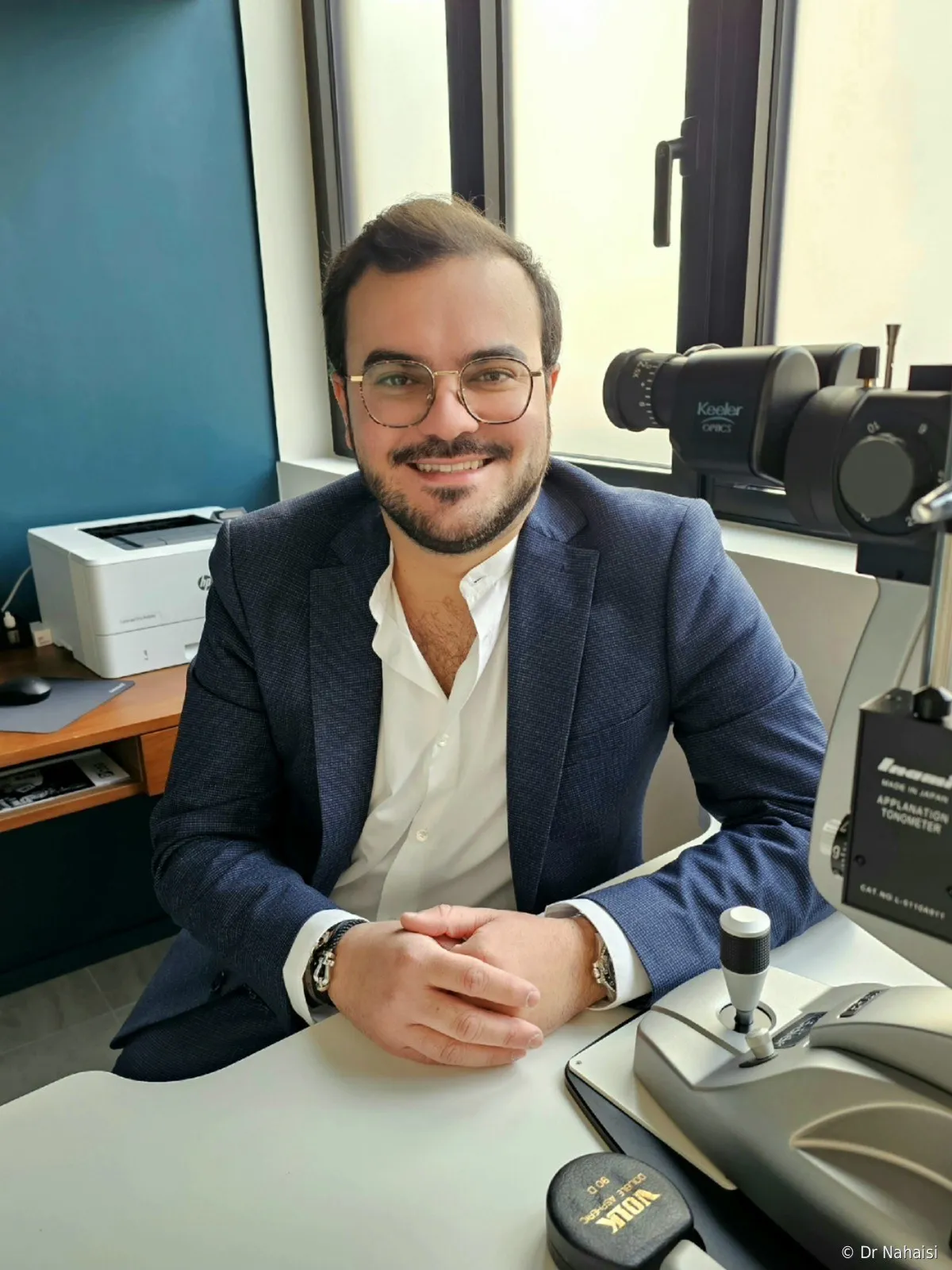
Dr Steven Nahaisi
Updated on 15 October 2026 · 35 min read
Refractive surgery is elective surgery. Nobody is obliged to have it. When an operation is not necessary, it is only right to explain its risks before you decide. During the consultation, I am not trying to confirm that an eye can be operated on; I am looking for the reason why it should not be. This page follows the same logic.
During the consultation. This is where risks are discussed, with test results in hand, before any decision.
On this page12 sections
- 02EXPECTED EFFECTS AND COMPLICATIONS
- 03EXPECTED EFFECTS
- 04COMPLICATIONS COMMON TO ALL TECHNIQUES
- 05COMPLICATIONS SPECIFIC TO EACH TECHNIQUE
- 06ECTASIA AND KERATOCONUS AFTER SURGERY
- 07CAN YOU GO BLIND?
- 08THE RISK OF NOT BEING SATISFIED
- 09WHAT I REFUSE TO DO
- 10WHEN SOMETHING IS WRONG
- 11THE SIGNS THAT SHOULD MAKE YOU CALL ME
- 12FAQ
- 13BOOK AN APPOINTMENT
EXPECTED EFFECTS AND COMPLICATIONS
Two different things that are often confused
Many of the messages I receive are about normal healing sensations. Here is the framework I use throughout this page.
Expected effects are part of the process. They are built into the biology of corneal healing, they follow a recognizable timeline and they subside. You get through them.
Complications are events that should not have happened. They require a diagnosis, treatment, sometimes a return to the operating room. They are rare, and most of my pre-operative work consists of preventing them from happening.
In between lies a gray area: an expected effect that lasts a very long time ends up becoming a problem. Three weeks of dryness is part of normal recovery; disabling dryness at one year is something else. The difference lies in duration and impact, not in the nature of the symptom.
EXPECTED EFFECTS
What almost everyone feels after laser surgery
Dry eye
This is the most common effect. Dryness is almost systematic at first, and it is temporary. It settles within a few weeks or a few months, with preservative-free artificial tears that you can use as often as you need.
The explanation is mechanical. The nerve trunks of the cornea enter from the periphery and run through the depth of the stroma, below the plane where the flap is cut. What the cut interrupts are the most superficial branches, at their front end. At the Institut Voltaire, we create thin flaps, around 100 microns, which further limits the tissue involved. These fibers regenerate completely in about 6 months, and the feeling of dryness disappears well before that. After TransPRK or SMILE, the nerves are less affected. In the end, there is slightly less dryness after TransPRK or SMILE, with no major difference.
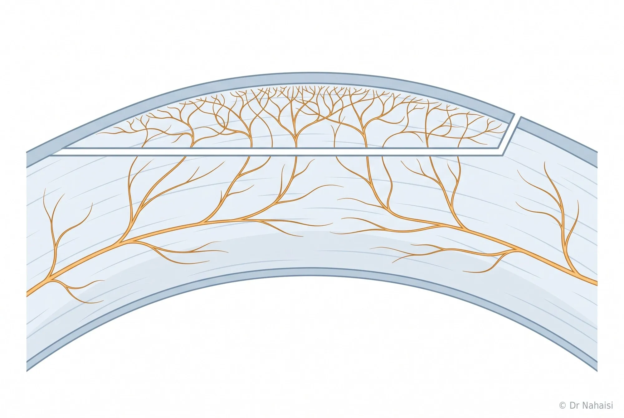
The nerves of the cornea, in cross-section. The trunks enter from the periphery and run through the depth of the stroma, below the plane where the flap is cut, where they remain intact. What the cut interrupts are their most superficial branches.
The pre-operative assessment includes an examination of your tear film, in quantity and quality. This is what allows me to tell you what to expect. When it is good, your dryness has no reason to be out of the ordinary or to last more than a few months. When it is fragile, we treat it before operating.
Fluctuating vision
Vision improves, dips a little, improves again, sometimes with differences between the two eyes. Everything evens out over about a month and stabilizes between 1 and 3 months. The full timeline is on the recovery page.
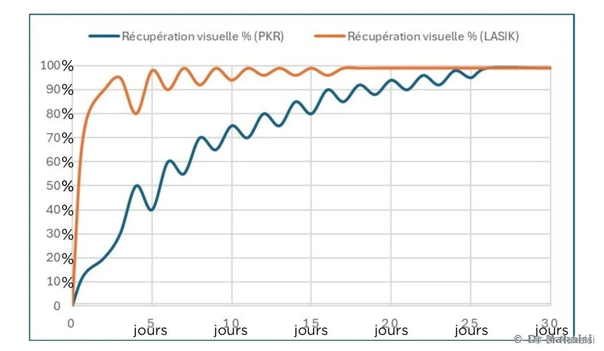
Visual recovery over 30 days, taken from my booklet. In orange, LASIK, which improves from the very next day. In blue, TransPRK, which is more gradual. The green curve shows the discomfort felt.
Recovery in detail, day by day
Halos and night vision
Halos around light sources, slightly spread-out headlights, glare in low light. These phenomena first increase, peaking around the second month, then gradually decrease, over 4 to 6 months for high corrections. Patients who had high astigmatism see many of them at first, because their refractive error was already producing them before surgery, and they are also the ones who notice the clearest improvementafterward. At 6 months and at one year, night vision is generally better than before the procedure.
With very high corrections combined with a large pupil, some discomfort may persist. I measure your pupil in low light during the assessment and I calibrate the optical zone so that it amply covers the dilated pupil. When your pupil is very large, I warn you before the procedure that part of the discomfort may remain. In everyday life, it is not very bothersome: the brain gradually tones it down over several months.
Eye strain and redness
In the first few days, the eye has trouble focusing up close, like a camera whose autofocus is recalibrating, sometimes with headaches at the end of the day in front of a screen. This gradually decreases over a month. If it persists, a small pair of resting glasses for screens solves the problem. It is not a failure of the surgery; it is a normal after-effect.
A red eye the next day, sometimes with a well-defined patch, was common with older machines. The Visumax 800 presses only on the cornea, and this is less pronounced, but it remains common and harmless. When it happens, the patch clears on its own in one to two weeks, like a bruise.
Discomfort in the first hours
After LASIK or SMILE, the eyes sting and water for 4 to 6 hours, then it settles down. After TransPRK, the discomfort lasts longer: a 48 to 72 hour phase, with real pain during the first two days. I come back to this below.
COMPLICATIONS COMMON TO ALL TECHNIQUES
What can happen, whatever the technique
Infection
This is the complication patients fear most, and it is extremely rare. Thank God, I have never had one in any of my patients. I would add something from my own background: I worked for five years at Cochin Hospital, three of them as head of the cornea department, and I never saw an infection there that followed refractive surgery. The infectious keratitis cases I treated came from contact lens wear, trauma, or vulnerable patients.
Surgery is performed under sterile conditions, and a topical antibiotic treatment is used as eye drops for two weeks after surgery. A corneal laser does not enter the eye: it stays at the surface, without opening the inside of the eye.
A corneal infection, when it occurs, can be treated: antibiotic eye drops, sometimes fortified preparations, sometimes a surgical washout of the interface after LASIK. Itshows up as pain that increases instead of decreasing, a red eye and a drop in vision, generally in the first week. You call me the same day.
Dryness that sets in
Cases of dryness persisting for several months are rare. I have had a few, and it has always eventually settled down, sometimes after six months, sometimes after a year.
When dryness sets in, I do not just prescribe artificial tears. There are enhanced tears, tear gels for the night, an anti-inflammatory eye drop, punctal plugs that keep tears in the eye, and an oral treatment that is useful in certain patients. Inserting the plugs takes a few seconds and does not hurt.
One mechanism is worth knowing about. Marked dryness is often accompanied by an inflammatory reaction that damages the surface of the cornea. This irregular surface diffracts light and creates astigmatism that can be measured at the check-up. It then feels as if the surgery has failed: it is the surface that needs treating, and vision returns once it is treated.
Undercorrection and overcorrection
The cornea heals, and healing retains an element of unpredictability. Sometimes part of the correction is missing, or the result slightly overshoots the target, generally by about half a diopter. This is more common with high corrections.
In my practice, it is rare. I operate on about 400 patients a year and I perform fewer than one enhancement a year in nearsighted and farsighted patients. It is a little more common after presbyopia correction, and it remains low.
The approach is always the same. We wait for stabilization, we measure objectively, and we decide. It is too early to talk about an enhancement before the end of the first six months, because part of the deviation corrects itself spontaneously. A simple pair of resting glasses is sometimes enough. Otherwise, we go as far as a laser enhancement.
Regression
The correction applied by the laser is lasting: the tissue removed does not grow back and the new curvature is maintained. What can change is the epithelium, the superficial layer of the cornea, which thickens again over the years and compensates for a small part of the ablation made beneath it. For a modest correction, this is on the order of 0.25 to 0.50 diopter over ten to fifteen years. A -1 nearsighted patient will almost never regress beyond -0.10 or -0.20 diopter, which has no visible effect on vision.
The higher the initial correction, the more regression is possible, particularly beyond -6 diopters of myopia, +4 of hyperopia or -2 of astigmatism. A -8 nearsighted patient who drifts back to -0.75 remains very happy with the result, and a laser enhancement is still possible if corneal thickness allows.
Two different mechanisms can bring back a small correction, and they are often confused.
Epithelial regression. This is the one I just described. The epithelium, the superficial layer of the cornea, thickens slightly again and offsets part of the laser's effect. It is the operated cornea that changes, not the eye. This phenomenon was significant fifteen or twenty years ago, with older generations of lasers. With the Schwind platform and current treatment profiles, it has become rare in simple and moderate myopia, and it remains moderate in high corrections.
Progression of the refractive error. Here, it is the eye itself that keeps changing, independently of the surgery, as if you had not been operated on and were changing glasses in a few years. For myopia, this mechanism is linked to the lengthening of the eye, which almost no longer occurs after age 23 or 24. An example: if you are at -2.00 today and your theoretical need were to go to -2.50 in ten years, you would be at -0.50 ten years after surgery. You willnot go back to your initial myopia.
That is why I look at the history of your eyeglass prescriptions during the assessment. And if the combination of the two brought back some discomfort after several years, an enhancement would still be possible in most cases.
Myopia: stability and progressionHyperopia and its laser correction
Loss of lines of corrected visual acuity
Your corrected visual acuity is the best you can see with the best possible correction, in glasses or contact lenses. It is the maximum your eye is capable of. Myopia, astigmatism or hyperopia do not change this maximum; they only prevent you from reaching it without correction.
The goal of surgery is to let you see without glasses what you used to see with them. A residual correction, regression or remaining astigmatism can be made up for with glasses or an enhancement. Losing lines of corrected acuity means that this maximum itself goes down, and no optical correction can make up for it.
This can happen after irregular healing, a corneal opacity, or a poorly managed complication. It is rare, and it is what the pre-operative assessment seeks to rule out first and foremost. A drop of one line during healing, on the other hand, is common and temporary.
Corneal ectasia
This is the most feared complication of LASIK. It has its own section, further down.
COMPLICATIONS SPECIFIC TO EACH TECHNIQUE
Each technique has its weak points
Each technique has its own risk profile, and this is one of the criteria I rely on to guide you, along with your cornea and your correction.
LASIK
LASIK has been the most widely performed refractive surgery in the world since the late 1990s. Its specific complications are almost all related to the existence of the corneal flap.
Difficulties in cutting the flap. The femtosecond laser creates the flap by placing thousands of micro-pulses side by side, each releasing a small gas bubble. With older generations, which were slower and used more energy, these bubbles could spread unevenly and leave small uncut areas. The Visumax 800 works at very low energy, with closely spaced pulses, and this phenomenon has practically disappeared. If a cut were incomplete, we put the flap back down, we do not treat, and we reschedule the procedure a few months later, generally as surface surgery. You have lost nothing.
Flap folds. The flap is laid back flat at the end of the procedure and adheres through natural suction, then through healing of its edge. Folds can appear, especially in the first hours and in case of rubbing. When they are central and visible, they impair the quality of vision, and we go back to the operating room to lift the flap again, rinse it and smooth it. Subtle micro-folds, on the other hand, are common and have no effect on vision.
Flap displacement. This is something extremely rare. The flap heals around its edge within a few hours, and this peripheral healing secures its position for the long term. The risk exists during the first 24 to 48 hours, in case of a direct blow or hard rubbing, and that is why you do not rub your eyes and you wear the eye shields for the first three nights after LASIK. Beyond that, ordinary rubbing no longer displaces it. A violent or very targeted trauma, however, can displace a flap even long after, several years after the procedure.
The cases found in the medical literature involve very targeted trauma, a cat scratch or a road accident, which would damage an eye that had never been operated on anyway. Prompt surgical repair then allows a perfect recovery in the vast majority of cases. This has happened to me once or twice out of several thousand patients operated on, and they always recovered well.
Diffuse lamellar keratitis. This is a sterile, non-infectious inflammatory reaction that develops between the flap and the underlying stroma in the first few days. It causes blurring, discomfort in light, sometimes nothing at all, and it is often me who spots it at the check-up. Mild forms, by far the most common, are treated with intensive topical corticosteroids and subside without after-effects. Cases that require lifting the flap again to rinse the interface are exceptional.
Epithelial ingrowth. Cells from the epithelium can migrate under the flap through the cut line and multiply in the interface. This is rare after a first surgery, clearly less so after the flap is lifted for an enhancement, and all the more so when this lift is done a long time later. That is one of the reasons why I often prefer to do enhancements on the surface. Peripheral forms remain stable and only require monitoring. Those that advance toward the center are treated by lifting the flap to clean the interface.
Everything about LASIK surgery in Paris
TransPRK
TransPRK is the safest technique from a biomechanical point of view: it does not create a flap and preserves more corneal thickness, so no flap displacement is possible, and the risk of ectasia is greatly reduced. Its risk profile is concentrated on surface healing.
Pain in the first 48 to 72 hours. Healing of the epithelium causes a foreign-body sensation, watering, strong sensitivity to light, and real pain during the first two days. The bandage contact lens reduces these sensations without eliminating them, and I systematically prescribe painkillers. How it feels varies from one patient to another, and some describe only stinging. It is a technique I have complete confidence in: I operated on my own wife with TransPRK.
Corneal haze. This is a slight opacity that can develop in the weeks following surface surgery, especially with high corrections. It is a healing reaction, encouraged by prolonged sun exposure, and the problem does not exist with LASIK, where the surface does not need to heal. It has become rare with TransPRK thanks to the antimitotic agent I systematically apply during the procedure, a soaked sponge placed on the cornea for about thirty seconds, with no added risk. When haze does appear despite this, it is most often asymptomatic, and treatment sometimes involves resuming corticosteroids. Advanced symptomatic forms have become practically nonexistent: I have not seen any since I began operating at the Institut Voltaire on recent machines, even though I perform TransPRK on myopia as high as -10.
Prevention comes down to one instruction: protect your eyes from the sun for the following six months. Well-tinted sunglasses are enough, wraparound ones in case of strong exposure at the seaside or in the mountains.
Delayed epithelial healing. The epithelium normally closes in 48 to 72 hours. In some patients, closure takes a little longer, in the context of dry eye or diabetes. The corneal epithelium constantly renews itself, in about a week, throughout life: a healthy cornea always heals in the end. A cornea that did not heal would be a cornea that was already diseased before surgery, with limbal stem cell deficiency, and that is something I check during the assessment.
Regression with high corrections. Surface healing is a little less predictable than LASIK healing for high corrections. An enhancement remains possible after complete stabilization.
Everything about TransPRK in Paris
SMILE
SMILE is available at the Institut Voltaire and I offer it when it is indicated. Its specific complications stem from the fact that the tissue removed must come out in a single piece through a micro-incision.
More complex enhancement. There is no flap to lift after SMILE. Enhancement is done either with TransPRK, with its 7 to 10 days of recovery, or by secondarily creating a flap for LASIK, depending on the case, never by creating a new lenticule.
Everything about SMILE in Paris
PresbyLASIK
The technical complications are those of LASIK, with the addition of adaptation difficulties specific to this surgery.
A poorly tolerated compromise. This is the real difficulty of PresbyLASIK. The brain has to learn to cope with two eyes that no longer see the same thing at the same distance. Most patients get used to it within a few weeks. Some describe discomfort at distance, a feeling of imbalance, fatigue. I reduce this risk by having you test the difference between the two eyes with contact lenses before surgery, when the strategy lends itself to it.
Reversibility. The Schwind platform makes it possible to go back: we adjust the profile, or we restore the cornea to its pre-treatment curvature.
ECTASIA AND KERATOCONUS AFTER SURGERY
The question that comes up most often
What ectasia is
Keratoconus is a progressive deformation of the cornea, which thins and bulges into a cone. It degrades vision irregularly, in a way that is difficult to correct with glasses. Post-operative ectasia is the same phenomenon, triggered or accelerated by surgery on a cornea that was predisposed or weakened too much. It is what every refractive surgeon seeks to avoid, because the treatment is heavy: rigid contact lenses, cross-linking, intracorneal rings, and in advanced forms, a corneal transplant.
A normal cornea, operated on according to the rules, does not become ectatic. Ectasia occurs in predisposed corneas that the assessment did not detect, in corneas too thin for the correction removed, or in the presence of chronic, repeated eye rubbing. I work on these three points.
First safeguard: pre-operative screening
The pre-operative assessment is not only used to check your correction. Above all, it is used to examine your cornea closely enough to identify those that should not be operated on.
Topography measures the curvature of your cornea point by point, on both of its surfaces. Advanced forms of keratoconus are obvious. Early forms, which do not yet cause any symptoms, require a trained eye: a subtle asymmetry between the two halves of the cornea, a localized thinning, a back surface that stands out.
Epithelial thickness mapping is my favorite test on this subject, and it is rarely performed in general ophthalmology. The epithelium, the superficial layer of the cornea, has a remarkable property: it compensates for irregularities in curvature. Over an area that is beginning to bulge, it thins to smooth out the relief, which masks the emerging deformation on curvature tests. Mapping it point by point amounts to removing this camouflage. A localized epithelial thinning over a bulging area is one of the earliest signs I have. This test is systematically part of the assessment at the Institut Voltaire, and it is what allows a decision on borderline corneas.
Added to this are pachymetry, mean and maximum keratometry, analysis of the back surface, and the indices calculated by topographers. There are dozens of formulas estimating the risk of post-operative ectasia, each giving more or less weight to one parameter or another, and none of them decides for me. My role is to review all these values comprehensively and to make a diagnosis of suitability for surgery. If it were just a matter of looking at corneal thickness or a score on a sheet, it would be a little too easy.
My training in corneal surgery, at the Hôtel-Dieu under Prof. Bourges, at the Fondation Rothschild with Prof. Gabison and Prof. Gatinel, then as Chief Resident (Chef de Clinique) at Cochin, taught me to spot these signs and to distinguish true positives from false positives. Refusing to operate on a patient who is perfectly suitable for surgery is also a mistake. When a case raises a question, I do not decide alone: at the Institut Voltaire, I work with Dr Camille Rambaud and Dr Romain Nicolau, who are also refractive surgeons, and we discuss borderline cases together every week.
Second safeguard: the residual stromal bed
The residual stromal bed is the thickness of intact cornea that remains beneath the treated area, and it is what guarantees the mechanical strength of your eye over decades. The calculation is simple and I do it for each eye. We start from the total thickness of your cornea, on average 525 microns. In LASIK, we subtract the thickness of the flap, on average 110 microns, sometimes reduced to 100 microns, or even 90 or 80 microns in very rare complex cases. We then subtract the ablation, about 15 microns per diopter of myopia corrected, a value that also depends on the diameter of the optical zone. What remains is the residual bed.
Traditional guidelines set the safety limit above 280 microns, and more recent work conducted in London has shown that a 250-micron bed in a regular cornea remained safe. I prefer a stricter approach and I set my limit at 300 microns, so that this risk is as low as possible.
The laser software simulates the ablation and displays the expected residual bed before each treatment. I check this value eye by eye, and when it falls below my limit, I change technique. TransPRK does not sacrifice a layer of stroma in a flap and preserves, for thesame correction, about sixty microns more. When that is not enough either, I do not operate with the laser.
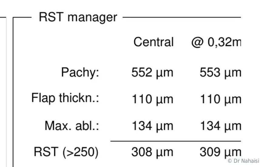
The “RST manager” of the Schwind laser, on an anonymized case. The software starts from the thickness of the cornea (552 microns), subtracts the thickness of the flap (110 microns) and the ablation depth (134 microns), and displays the stromal bed that will remain: 308 microns. It is this calculation, done for each eye, that rules out the risk of ectasia.
Third safeguard: eye rubbing
This is the most underestimated factor, and it is the one you have control over.
There is a predisposition to corneal deformation, which can be hereditary. But there is no progression to keratoconus in the absence of rubbing. This is the “no rub, no cone” theory, championed by Prof. Gatinel, one of my mentors. The gesture is not always conscious: rubbing on waking up, allergic itching, sleeping on your stomach with your head in the pillow, a hand or elbow under the eye during the night, rubbing in front of screens. What matters is strong pressure applied to the eyeball. Gently running your fingers around the eyes is not a problem, preferably on the bony rim of the eye socket, and this is the habit I teach my patients to adopt after the procedure.
When topography shows a sign of fragility, I look for a habit of this kind, and it then becomes a topic of consultation in its own right. Identifying and changing it is sometimes enough to stabilize a fragile cornea and to make surgery possible after a few months of follow-up. When the itching comes from an allergy, antihistamine treatment often solves the problem.
After surgery, I recommend that all my patients limit rubbing for good. It is general eye hygiene advice, and I also give it to people I do not operate on.
And cross-linking
Cross-linking strengthens the bonds of corneal collagen and is used to stabilize progressive keratoconus.
I reserve it for certain cases, especially in young patients, under 25. In a teenager who will not follow the instructions about rubbing, the indication seems solid to me. In an adult who has understood the mechanism and who really stops rubbing their eyes, I am more reserved, because keratoconus rarely progresses once rubbing stops. If progression is demonstrated nonetheless, the indication becomes legitimate.
What this means in practice
A thin but regular cornea, with a small correction to remove, keeps a comfortable residual bed and can be operated on with LASIK. A thick cornea that shows an unexplained asymmetry will be steered toward surface surgery, or not operated on. A regular cornea at 490 microns can be more suitable for LASIK than an irregular cornea at 520 microns.
CAN YOU GO BLIND?
The answer is no
I would say that you cannot go blind from refractive surgery performed under normal conditions, with a favorable pre-operative assessment.
The explanation is anatomical. Vision originates at the back of the eye, in the retina, and the information travels to the brain through the optic nerve. These two structures carry the risk of blindness, and they are the ones affected in the diseases that cause blindness: advanced glaucoma, macular degeneration, retinal vascular occlusions, extensive retinal detachments.
The refractive laser never goes there. It works on the cornea, to a depth of a few tens to a few hundred microns. It does not touch the lens, the vitreous, the retina or the optic nerve, and no internal structure of the eye is opened.
Serious complications do exist, an infection for example. They are extremely rare, and they can be treated.
What is possible, and I do not minimize it, is a deterioration in the quality of vision: a drop in corrected acuity, persistent light phenomena, significant dryness. These situations are rare, and it is these, not blindness, that constitute the real risk of this surgery.
If this worry persists, we can talk on the phone, or you can come back to see me for a consultation to go over all your questions, free of charge. Some patients leave without having surgery, and that is a valid answer.
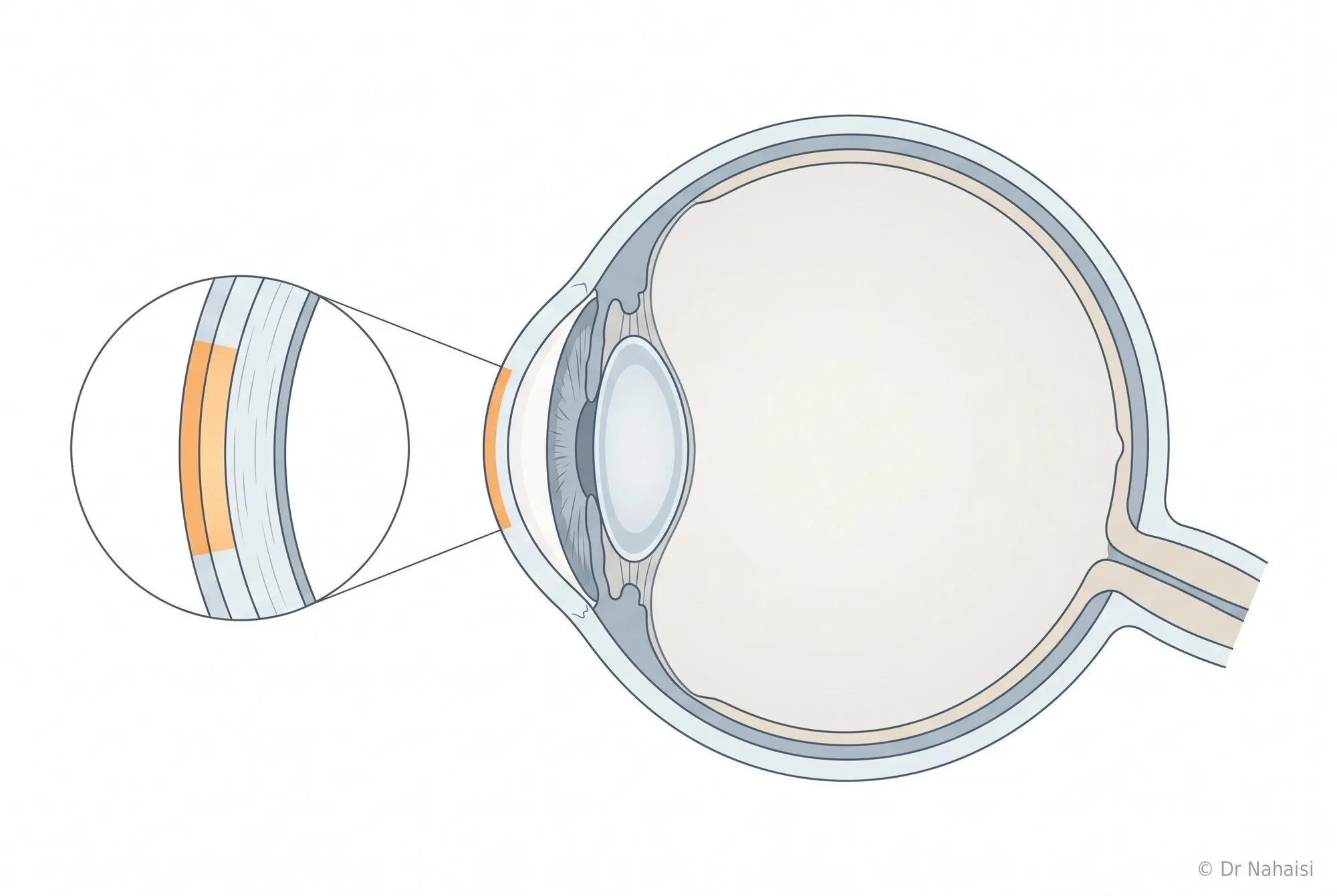
The eye in cross-section. In orange, the area where the laser works: the cornea, to a depth of a few tens to a few hundred microns. The lens, the retina and the optic nerve, which are the structures whose damage can lead to blindness, are never touched.
THE RISK OF NOT BEING SATISFIED
A successful surgery and a disappointed patient
It happens: a flawless cornea, an excellent measured acuity, and someone who is not happy. This situation does not appear on any list of complications, and it deserves to be dealt with like the others.
Expectations that do not match what the technique can achieve
The mismatch happens before the surgery. A 45-year-old nearsighted patient who comes in to never wear glasses again will be disappointed if their distance vision is corrected without discussing their presbyopia. Someone who hopes for perfect vision in all circumstances, day and night, without the slightest halo, will find the first weeks long.
During the assessment, I compare what you expect with what surgery can achieve for your eyes, and I tell you when the gap is too wide.
The compromise for presbyopic patients
Correcting presbyopia with laser means giving something up. With monovision and blended vision, one eye sees well at distance, the other sees well up close, and the brain combines the two. With a multifocal profile, each eye spreads light over several distances. In both cases, we do not recreate the vision of a 25-year-old: we trade a little distance comfort for near-vision independence.
Most patients find the trade largely worthwhile, but in rare cases some do not adapt. I have nearsighted patients test the compromise with contact lenses before surgery, when the strategy lends itself to it. A pair of occasional glasses remains useful for demanding tasks, subtitles at the movies or prolonged reading. When the compromise still does not suit, we can intervene again after a stabilization period of at least 6 months. This enhancement ranges from a simple readjustment to a complete reversal of the treatment; that is the advantage of Schwind's PresbyMAX platform.
Patients for whom I think the result will be disappointing
I sometimes refuse to operate on someone whose eyes are perfectly suitable for surgery. A patient with very mild myopia who hopes for a radical change will be disappointed by an improvement their eyes cannot produce. Someone whose main complaint is eye strain due to an unsuitable correction does not need laser surgery; they need good glasses. Someone who comes reluctantly, pushed by those around them, has nothing to gain from having surgery now. In these cases, I say no, or I suggest waiting.
What I do when a patient is not happy
I see them again, as many times as necessary, free of charge, and I measure objectively. A complaint compared against a refraction, a topography and an examination of the surface almost always yields an explanation. In many of these situations, the cause is not the laser treatment itself: it is dryness affecting the surface, accommodative fatigue that resolves with resting glasses, residual astigmatism that is still regressing, a slower-than-average recovery. Each has a solution, and none requires going back to the operating room. When a repeat procedure is justified, we do it, but not before the end of the first six months.
WHAT I REFUSE TO DO
My red lines
I prefer not to operate rather than take a risk with your eyes. Here is what that covers.
I do not operate on a questionable cornea. Confirmed keratoconus, serious suspicion, an asymmetry I cannot explain: I hold off and see the patient again a few months later with new topographies.
I do not operate when the calculated residual bed falls below 300 microns. My limit is stricter than standard guidelines, and I do not move it.
I do not operate on a correction that has not stabilized. This is most common between ages 18 and 24. We wait six months or a year, we repeat comparable measurements, and often the answer becomes yes. I do still sometimes operate on 18-year-old patients, warning them that progression remains possible.
I do not operate on an eye surface in poor condition. Severe dryness, blepharitis, poorly controlled allergy: we treat first, then reassess. Many patients come back a few months later in much better condition.
I do not operate during pregnancy, as a precaution. Breastfeeding, on the other hand, is not a problem at all, and there is no need to wait until you have had all your children.
I do not operate on someone who I think will be disappointed by the result. This is the hardest line to hold, because it relies on judgment.
I do not operate beyond the limits of the laser. LASIK corrects myopia up to -10 diopters, sometimes -12, hyperopia up to +7 or +8, astigmatism up to -6. Beyond that, and for thin or deformed corneas, other solutions can sometimes be considered, and I refer patients to colleagues.
A no today is not always a no forever. An unstabilized correction, a cornea weakened by rubbing, a surface that needs treatment, an ongoing pregnancy are temporary situations. Confirmed keratoconus or a structurally too-thin cornea will not change. I always tell you which category your case falls into.
WHEN SOMETHING IS WRONG
What happens, in practice
You can reach me
Phone and WhatsApp:01 84 80 28 04. By email:nahaisi@laservisionparis.com. Or through Doctolib messaging.
I answer personally, including in the evening and on weekends. WhatsApp is often the easiest, and a photo of the eye sometimes saves you a trip across Paris.
I see you again
Post-operative follow-up is unlimited and included in the fee for the procedure. You come back as many times as necessary, including years later. The first check-up takes place on the Monday afternoon following the Friday surgery, sometimes on Wednesday or Thursday.
We can often sort things out remotely. When it persists, I prefer to see you within the following days.
I perform a repeat procedure if necessary
An enhancement is possible in most cases, after complete stabilization. After LASIK, by lifting the existing flap and readjusting with the excimer laser. After TransPRK, on the surface. After SMILE, with TransPRK or by secondarily creating a LASIK flap, depending on the case, never by creating a new lenticule.
The enhancement is included in the initial fee for the first year, then offered at a preferential rate up to 5 years. If urgent action is needed, a flap to reposition for example, we go back to the operating room without delay and without any discussion of fees.
I do not leave you alone with a problem
When a situation calls for a second opinion, I say so and I arrange it. I discuss it with my colleagues at the Institut Voltaire, I refer you to a specialist when necessary, and I continue to follow your case.
Fees, quotes and complementary health insurance reimbursement
THE SIGNS THAT SHOULD MAKE YOU CALL ME
What is normal, and what is not
| Normal, you can wait for the check-up | Call me |
|---|---|
| Eyes that sting and water in the first hours | Pain that increases instead of decreasing after the 3rd day |
| Blurry or veiled vision in the first few days | A sudden drop in vision in one eye, when things were getting better |
| Vision that fluctuates from one day to the next | Red eye with pain and light sensitivity that are getting worse |
| Red white of the eye, looking like burst blood vessels | A feeling that you took a knock or rubbed your eye hard in the first 48 hours |
| Halos around lights in the evening | Thick discharge or eyelid stuck shut on waking, with pain |
| Gritty, sand-like sensation, dryness | Bandage contact lens fallen out or wrinkled before the check-up, after TransPRK |
| Difference between the two eyes for several weeks | A clear impression that the veil is coming back after having disappeared |
| Eye strain and headaches at the end of the day | Any question this page does not answer |
Phone and WhatsApp:01 84 80 28 04. By email:nahaisi@laservisionparis.com. Or through Doctolib messaging.
Frequently asked questions
Your questions about the risks of refractive surgery
Can you go blind after laser eye surgery?
I would say no: you cannot go blind from refractive surgery performed under normal conditions, with a favorable pre-operative assessment. The laser works only on the cornea. It does not touch the lens, the retina or the optic nerve, which are the structures whose damage can lead to blindness. Serious complications exist; they are extremely rare and they can be treated.
What is the risk of infection after LASIK?
It is extremely rare. Thank God, I have never had one in any of my patients. I also worked for five years at Cochin Hospital, three of them as head of the cornea department, and I never saw an infection there that followed refractive surgery. Surgery is performed under sterile conditions and a topical antibiotic treatment is used as eye drops for two weeks. An infection, if it occurred, can be treated.
What is corneal ectasia and what is the risk after LASIK?
Ectasia is a progressive deformation of the cornea, which thins and bulges, as in keratoconus. It occurs in corneas that are predisposed or weakened too much, not in normal corneas operated on according to the rules. Three things prevent it: pre-operative screening, a sufficient residual stromal bed, and stopping eye rubbing.
Can LASIK trigger keratoconus?
In a patient with no predisposition, no, except with an unsuitable treatment. In a patient with undetected early keratoconus, LASIK can make it worse, because it removes thickness from an already fragile cornea. Everything depends on screening: topography of both surfaces and epithelial thickness mapping detect the forms that do not yet cause any symptoms. When a cornea is suspicious without certainty, I systematically avoid LASIK.
What is the residual stromal bed after refractive surgery?
It is the thickness of intact cornea left beneath the treated area, and it is what guarantees the mechanical strength of the eye over the long term. Traditional guidelines place it above 280 microns. I set myself 300 microns, I check this value eye by eye before each surgery, and I change technique when it is not reached.
Can the LASIK flap move?
It is extremely rare. The flap heals around its edge within a few hours, and this healing secures its position. The risk exists during the first 24 to 48 hours, in case of a direct blow or hard rubbing, which is why the eye shields are worn for the first three nights. Beyond that, ordinary rubbing no longer displaces it. The cases described in the medical literatureinvolve very targeted trauma, and prompt repair then allows a perfect recovery.
Can dry eye after LASIK last forever?
It does not persist long-term. It is almost systematic at first and temporary. Cases lasting several months are rare: I have had a few, and it has always eventually settled down, sometimes after six months, sometimes after a year. Corneal nerve fibers regenerate in 6 months on average.
Can halos after laser eye surgery last for life?
They first increase, peaking around the second month, then decrease, over 4 to 6 months for high corrections. At 6 months and at one year, night vision is generally better than before the procedure. Minimal discomfort may persist with very high corrections combined with a large pupil, and that is why I measure your pupil during the assessment.
Can you lose lines of vision after refractive surgery?
A temporary drop of one line during healing is common. A lasting loss of corrected acuity, that is, of the best you can see even with glasses, is a rare complication. It can follow irregular healing, a corneal opacity, or a poorly managed complication. This is the risk the pre-operative assessment targets first and foremost.
What is haze after TransPRK?
It is a slight opacity of the cornea, a healing reaction that can develop after surface surgery, especially with high corrections and after prolonged sun exposure. This problem does not exist with LASIK. It has become rare thanks to the antimitotic agent systematically applied during the procedure and to improvements in lasers, and when it does appear it is most often asymptomatic. Prevention consists of protecting your eyes from the sun for six months.
Is pain after TransPRK a complication?
No, it is an expected after-effect. Healing of the epithelium causes real discomfort for 48 to 72 hours, with real pain during the first two days. The bandage contact lens and the painkillers I systematically prescribe help you get through this phase, and some patients feel only stinging.
Does SMILE have any particular risks?
Its specific complication is difficulty extracting the lenticule, the small disc of tissue that must come out whole through a micro-incision. It can tear or leave a fragment behind, which lengthens the procedure. The Visumax 800 separates it much more cleanly than previous generations. The other point to know is enhancement, which is done with TransPRK, never by creating a new lenticule.
What happens if I cannot tolerate the PresbyLASIK compromise?
We intervene again, after a stabilization period of at least 6 months: adjustment of the profile, or a complete return to the pre-treatment curvature, as the Schwind platform allows this reversibility. Beforehand, I have you test the compromise with contact lenses when the strategy lends itself to it.
How long do you have to wait before an enhancement after refractive surgery?
I never perform an enhancement before the end of the first six months. Healing is still at work throughout this time, and things most often improve on their own. The enhancement is included in the first year, then offered at a preferential rate up to 5 years.
I am worried about having laser eye surgery. What should I do?
Come and talk about it. You can come back to see me for a consultation to go over all your questions, free of charge, or write to me. The most important thing with this kind of surgery is to feel ready, however long that takes, and there is no urgency to operate. If it is LASIK that worries you, TransPRK avoids any flap-related complication, since it does not create one, and greatly reduces the risk of ectasia.
BOOK AN APPOINTMENT
A consultation to ask all your questions
You do not need a referral letter, a prescription, or a prior visit to another ophthalmologist to come and see me. The consultation is at the standard rate set by French national health insurance with no fee supplement at my practices in the 10th and 19th, with nothing to pay upfront (direct billing). At the Institut Voltaire, in the 11th, you pay €50 upfront, including a €30 fee supplement that your complementary health insurance reimburses at least €20 of, and often in full.
Come with your questions, including the ones that seem silly to you.
Where I see patients
- Institut Laser Ophtalmologique Voltaire: 5 rue La Vacquerie, 75011 Paris. Consultations on Wednesday afternoons, surgery on Friday mornings, sometimes on Wednesday afternoons. Metro Voltaire (line 9), Philippe Auguste (line 2).
- 19th arrondissement practice: 37 rue Archereau, 75019 Paris. Consultations on Monday afternoons. Metro Crimée (line 7).
- 10th arrondissement practice: 12 rue du Faubourg Saint-Martin, 75010 Paris. Consultations on Thursday afternoons. Metro Strasbourg Saint-Denis (lines 4, 8, 9).
Book an appointment on Doctolib
Phone and WhatsApp:01 84 80 28 04. By email:nahaisi@laservisionparis.com. Or through Doctolib messaging.
Download the information booklet (PDF)
The pre-operative assessment in detail
“I don’t operate on eyes, I operate on patients.”
Dr Steven Nahaisi, ophthalmic surgeon in Paris, specialist in laser refractive surgery.
Author: Dr Steven Nahaisi, ophthalmic surgeon, specialist in laser refractive surgery, Institut Laser Ophtalmologique Voltaire, Paris 11th.
Your surgeon
Dr Steven Nahaisi
Ophthalmic surgeon dedicated exclusively to laser refractive surgery. He operates at the Institut Laser Ophtalmologique Voltaire and consults in three Paris offices. Consultations in English.