
Case study
Can high hyperopia be treated with laser eye surgery? A case at +5
Yes, high hyperopia can be treated with laser eye surgery. Here is the case of a patient at +5 treated with LASIK: assessment, filmed surgery and results at 3 months.
Dr Steven Nahaisi, ophthalmic surgeon · October 27, 2026 · 9 min read
“Doctor, can hyperopia be treated with laser surgery?” Many patients ask me this, often with one idea in mind: laser eye surgery would only be for nearsighted people. For farsightedness, there would be nothing, or almost nothing, to be done.
That was partly true twenty years ago. It is no longer the case. Today’s lasers correct every type of refractive error, and hyperopia is one of them. Some studies report encouraging results up to +8 diopters in carefully selected patients [4, 5]. What is actually possible depends on the cornea, the optical profile and the laser platform used.
Why hyperopia long had a bad reputation
The first lasers were designed for myopia. For hyperopia, their capabilities were much more limited. In the United States, the first wavefront-guided custom treatment approved by the FDA for hyperopia stopped at +3 diopters; other lasers of the time went up to +6, but with accuracy that dropped beyond +4 [6]. Correcting high hyperopia sometimes took two successive treatments.
Current platforms have changed this: finer beams, faster eye-tracking systems, wider and better-calculated treatment profiles. Hyperopia of +5 or +6 is now corrected in a single procedure, with very good results [1, 2].
The following case is a good example. My patient agreed that I could share his file, his examinations and the videos of his surgery, after anonymization.
The patient: 26 years old, +5 in both eyes
A sporty young man with no eye health problems. His hyperopia is high and broadly stable: +4.75 in the right eye and +4.50 in the left eye in his glasses. He tolerates contact lenses well but only wears them one day a week. The rest of the time, he wears glasses with thick lenses and a magnifying effect that enlarges his eyes and bothers him.
His request is simple: no longer depend on his glasses.
The assessment: what changes for a hyperope
The assessment includes most of the usual pre-operative assessment. But hyperopia has a few specific features, which come from the way the laser corrects it.
For myopia, the laser flattens the center of the cornea. For hyperopia, it is the opposite: the cornea must be made steeper. The laser therefore works in a ring around the center, thinning the cornea at the periphery: the cornea thus becomes steeper in the center, and therefore more powerful (see how laser eye surgery works). Three points then need particular attention.
Measuring the true hyperopia. A young hyperope constantly makes an effort to see clearly: this is accommodation. This effort hides part of the error. The correction is therefore measured after drops that relax accommodation (cycloplegia), or with the aberrometer. In his case, the measurements agreed: +5.00 (-0.50 at 165°) in the right eye and +4.50 (-0.25 at 20°) in the left eye, with 20/20 vision in both eyes with glasses.
Predicting the final corneal curvature. This is the key point. Each diopter corrected steepens the cornea by about one diopter. His cornea measured 43.4 diopters: the estimated final curvature was close to 48 diopters, a value compatible with our surgical goal. A limit of around 49 to 50 diopters has traditionally been used, beyond which quality of vision was thought to deteriorate. A recent study of more than 1,700 eyes questions this threshold: beyond 49 diopters, vision was not worse once other factors were taken into account [8]. There is therefore no absolute threshold that applies to every patient. As a matter of principle, however, I prefer to hold myself to this standard and stay below it.
Taking the pupil into account. The treatment must cover a zone wider than the pupil at night, to limit halos. His pupils measured 6.4 mm on the right and 6.1 mm on the left in the dark.
The rest of the assessment was reassuring. The Pentacam showed a thick cornea, 570 microns at the center, regular on both surfaces, with no sign of keratoconus: the screening index was 0.93, well within the normal range.
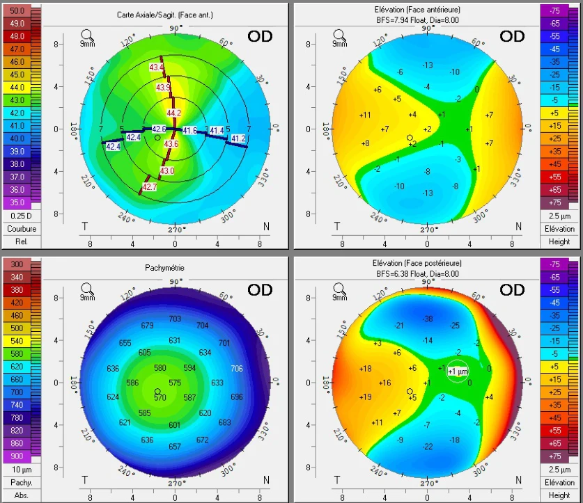
The same examination showed a shallow anterior chamber, 2.54 mm, which is usual in hyperopes, whose eyes are shorter. This is also why an implant is rarely a good option for hyperopia: there is often not enough room to place it.
Finally, aberrometry measures the correction point by point across the whole pupil. In the right eye, it ranged from +3.4 to +5.3 depending on the zone, with very few irregularities. The measured astigmatism matched that of his glasses, so I included it in the treatment.
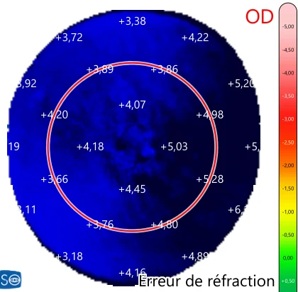
In summary: a thick, regular cornea and a final curvature that was predictable and within safety limits. LASIK was the right indication, in both eyes.
The treatment plan
The treatment was calculated on the Schwind Amaris laser, with an aspheric profile designed to limit aberrations. For the right eye:
- a 6.70 mm optical zone, wider than the pupil at night, extended by a 2 mm transition zone, for a total treated diameter of 8.70 mm;
- a ring-shaped ablation, almost zero at the center, maximal at the periphery;
- a 120-micron flap;
- a target curvature of 48.06 diopters.
What about corneal thickness? In myopia, thickness is often the limiting factor: the laser removes tissue at the center, where the cornea is thinnest. In hyperopia, it is the opposite. At the center, the laser removes almost nothing, barely 1 micron: with 570 microns of thickness and a 120-micron flap, about 450 microns of intact stroma remained. Tissue removal is greatest at the periphery, 108 microns on a circle about 7.2 mm in diameter. But the cornea is naturally thicker there, 687 microns: 459 microns still remained under the flap and the treated zone. This is why thickness calculations are far less restrictive in hyperopia than in myopia.
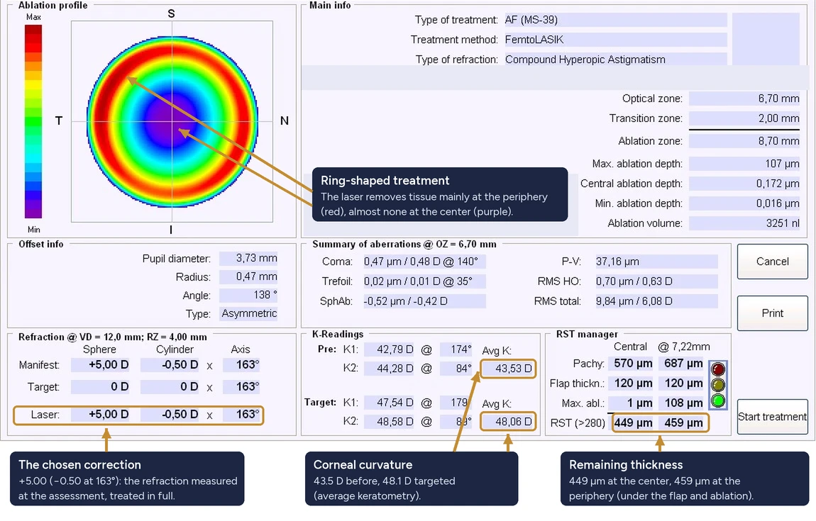
Because the treated zone is wide, the flap must be wide too, to fully cover the ring.
The surgery, on video
Both eyes were operated on the same day, under simple anesthetic drops. The videos are subtitled, step by step.
Step one: cutting the flap with the femtosecond laser (Visumax 800).
Step two: reshaping the cornea with the excimer laser (Schwind Amaris). The flap is marked and lifted, then the laser reshapes the cornea while a system continuously follows the movements of the eye. For the left eye, the laser treatment lasted 21 seconds: it takes a little longer for hyperopia, about 4 seconds per diopter, compared with 3 for myopia. The flap is then laid back, rinsed and smoothed.
The results
Five days later. 9/10 (about 20/22) in each eye without correction, 20/20 with both eyes, and perfect near vision. When measured, both eyes were slightly nearsighted, around -0.50. After LASIK for hyperopia, a slight, temporary myopic overcorrection may be observed. It generally decreases over the first few weeks, but how it evolves varies from patient to patient. I always explain this before surgery, so that this slight distance blur in the first days is not a worry.
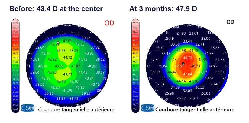
Three months later. 20/20 in each eye and 20/13 with both eyes for distance, with excellent near vision without correction as well. The right cornea measured 47.9 diopters, for 48.06 targeted. The left eye kept a very slight hyperopia when measured, with no effect on vision. This is what we aim for in these treatments: it is better to remain very slightly hyperopic than to become nearsighted.
What the epithelium does after surgery
The epithelium, the thin surface layer of the cornea, does not stay passive. At three months, it had thinned in the center and thickened in a ring around the treated zone. This is a well-known phenomenon after LASIK for hyperopia [3]: the epithelium partly smooths out the shape created by the laser.
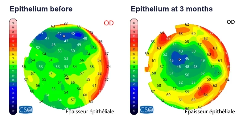
This explains why partial regression of the correction can occur over months or years, particularly in high hyperopia. Its extent varies from patient to patient. In a young patient, the eye usually compensates without difficulty. If it becomes bothersome, light computer glasses or an enhancement can be discussed. This is also why I follow these patients for longer (see recovery after surgery).
What the studies say
This result is nothing exceptional, which is rather good news. A Paris team followed 117 hyperopic eyes, at +5.1 on average, treated with LASIK using an adapted profile: at one year, the mean correction had returned to zero and 88% of eyes saw 20/20 without glasses [1]. With an older generation of Schwind laser (the Esiris), another series already found good results in hyperopia up to +7 [2]. Larger studies, up to +8, confirm the effectiveness and safety of LASIK for hyperopia, with appropriate follow-up [4, 5]. In 2026, a review by the American Academy of Ophthalmology, covering twenty studies, concluded that LASIK for hyperopia performed with current lasers offers high rates of effectiveness, predictability, safety and stability. Stability was lower for the highest corrections, older lasers and small optical zones [7].
Key points
- Yes, high hyperopia can be treated with laser eye surgery. Encouraging results are reported up to +8 in carefully selected patients, depending on the cornea and the laser platform.
- The key point of the assessment is the final curvature of the cornea. There is no absolute threshold, but I prefer to keep it below 49 to 50 diopters.
- The laser works in a ring, over a wide zone, adapted to the size of the pupil.
- Slight distance blur in the first days is possible: it is a small, temporary overcorrection that generally decreases within a few weeks.
- After high corrections, partial regression is possible over time; it is monitored and treated if needed.
Every cornea is different, and it is the assessment that shows what is possible in your case. To get a first idea, you can also answer the Am I a candidate? questionnaire.
Frequently asked questions
Can high hyperopia be corrected with LASIK?
Yes, within the limits of the cornea. Some studies report encouraging results up to +8 diopters in carefully selected patients. The deciding factor is the curvature of the cornea after treatment: there is no absolute threshold, but I prefer to keep it below 49 to 50 diopters.
Why is distance vision slightly blurred just after LASIK for hyperopia?
Because a slight, temporary myopic overcorrection may be observed: the eye is a little nearsighted in the first days. It generally decreases over the first few weeks, but how it evolves varies from patient to patient.
Can hyperopia come back after LASIK?
Partial regression is possible after high corrections, mainly because the epithelium thickens around the treated zone. Its extent varies from patient to patient, which is why longer follow-up is needed. Light computer glasses or an enhancement can be discussed if needed.
Sources
- Salah-Mabed I, Debellemanière G, Debellemanière G, et al. High hyperopic LASIK with reduction of corneal prolateness to control-induced spherical aberration. J Cataract Refract Surg. 2024;50(9):919-925.
- Alió J, Galal A, Ayala MJ, Artola A. Hyperopic LASIK with Esiris/Schwind technology. J Refract Surg. 2006;22(8):772-781.
- Reinstein DZ, Archer TJ, Gobbe M, et al. Epithelial thickness after hyperopic LASIK: three-dimensional display with Artemis very high-frequency digital ultrasound. J Refract Surg. 2010;26(8):555-564.
- Reinstein DZ, Carp GI, Archer TJ, et al. LASIK for the correction of high hyperopic astigmatism with epithelial thickness monitoring. J Refract Surg. 2017;33(5):314-321.
- Varley GA, Huang D, Rapuano CJ, et al. LASIK for hyperopia, hyperopic astigmatism, and mixed astigmatism: a report by the American Academy of Ophthalmology. Ophthalmology. 2004;111(8):1604-1617.
- U.S. Food and Drug Administration. List of FDA-Approved Lasers for LASIK.
- Santhiago MR, Steigleman WA, Al-Mohtaseb ZN, et al. LASIK for Hyperopia: A Report by the American Academy of Ophthalmology. Ophthalmology. 2026;133(4):466-476.
- Sella R, Sorkin N, Safir M, et al. Hyperopic LASIK and postoperative corneal steepness: revisiting the 49-diopter limit. J Cataract Refract Surg. 2024;50(6):550-557.
Read next
A question about your own eyes? The simplest way is to discuss it in consultation.
Book an appointment